4RG4

Epsilon-caprolactone-bound crystal structure of cyclohexanone monooxygenase in the Loose conformation


Experimental Data Snapshot

  • Method: X-RAY DIFFRACTION
  • Resolution: 2.51 Å
  • R-Value Free: 0.264 
  • R-Value Work: 0.182 
  • R-Value Observed: 0.191 

wwPDB Validation   3D Report Full Report


Ligand Structure Quality Assessment 


This is version 1.3 of the entry. See complete history


Literature

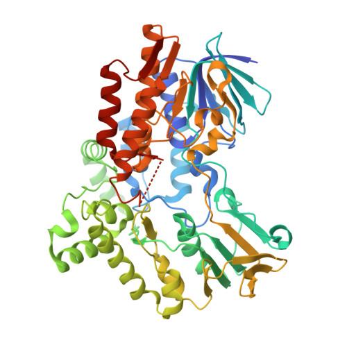
Lactone-bound structures of cyclohexanone monooxygenase provide insight into the stereochemistry of catalysis.

Yachnin, B.J.McEvoy, M.B.MacCuish, R.J.Morley, K.L.Lau, P.C.Berghuis, A.M.

(2014) ACS Chem Biol 9: 2843-2851

  • DOI: https://doi.org/10.1021/cb500442e
  • Primary Citation of Related Structures:  
    4RG3, 4RG4

  • PubMed Abstract: 

    The Baeyer-Villiger monooxygenases (BVMOs) are microbial enzymes that catalyze the synthetically useful Baeyer-Villiger oxidation reaction. The available BVMO crystal structures all lack a substrate or product bound in a position that would determine the substrate specificity and stereospecificity of the enzyme. Here, we report two crystal structures of cyclohexanone monooxygenase (CHMO) with its product, ε-caprolactone, bound: the CHMO(Tight) and CHMO(Loose) structures. The CHMO(Tight) structure represents the enzyme state in which substrate acceptance and stereospecificity is determined, providing a foundation for engineering BVMOs with altered substrate spectra and/or stereospecificity. The CHMO(Loose) structure is the first structure where the product is solvent accessible. This structure represents the enzyme state upon binding and release of the substrate and product. In addition, the role of the invariant Arg329 in chaperoning the substrate/product during the catalytic cycle is highlighted. Overall, these data provide a structural framework for the engineering of BVMOs with altered substrate spectra and/or stereospecificity.


  • Organizational Affiliation

    Departments of †Biochemistry and ‡Microbiology & Immunology, McGill University , 3649 Promenade Sir William Osler, Bellini Pavilion, Room 466, Montreal, Quebec, Canada H3G 0B1.


Macromolecules
Find similar proteins by:  (by identity cutoff)  |  3D Structure
Entity ID: 1
MoleculeChains Sequence LengthOrganismDetailsImage
Cyclohexanone monooxygenase548Rhodococcus sp. HI-31Mutation(s): 0 
Gene Names: chnBchnB1
EC: 1.14.13.22
UniProt
Find proteins for C0STX7 (Rhodococcus sp. HI-31)
Explore C0STX7 
Go to UniProtKB:  C0STX7
Entity Groups  
Sequence Clusters30% Identity50% Identity70% Identity90% Identity95% Identity100% Identity
UniProt GroupC0STX7
Sequence Annotations
Expand
  • Reference Sequence
Experimental Data & Validation

Experimental Data

  • Method: X-RAY DIFFRACTION
  • Resolution: 2.51 Å
  • R-Value Free: 0.264 
  • R-Value Work: 0.182 
  • R-Value Observed: 0.191 
  • Space Group: P 21 21 21
Unit Cell:
Length ( Å )Angle ( ˚ )
a = 55.145α = 90
b = 67.028β = 90
c = 133.576γ = 90
Software Package:
Software NamePurpose
DENZOdata reduction
SCALEPACKdata scaling
REFMACrefinement
PDB_EXTRACTdata extraction
StructureStudiodata collection
HKL-2000data reduction
REFMACphasing

Structure Validation

View Full Validation Report


Ligand Structure Quality Assessment 


Entry History 

Deposition Data

Revision History  (Full details and data files)

  • Version 1.0: 2014-10-15
    Type: Initial release
  • Version 1.1: 2015-01-14
    Changes: Database references
  • Version 1.2: 2017-11-22
    Changes: Refinement description
  • Version 1.3: 2023-09-20
    Changes: Data collection, Database references, Derived calculations, Refinement description