4RDA

X-RAY STRUCTURE OF THE AMYLOID PRECURSOR PROTEIN-LIKE PROTEIN 1 (APLP1) E2 DOMAIN IN COMPLEX WITH A HEPARIN DODECASACCHARIDE


Experimental Data Snapshot

  • Method: X-RAY DIFFRACTION
  • Resolution: 2.50 Å
  • R-Value Free: 0.245 
  • R-Value Work: 0.216 
  • R-Value Observed: 0.218 

wwPDB Validation   3D Report Full Report


This is version 2.1 of the entry. See complete history


Literature

Interaction of the amyloid precursor protein-like protein 1 (APLP1) E2 domain with heparan sulfate involves two distinct binding modes.

Dahms, S.O.Mayer, M.C.Roeser, D.Multhaup, G.Than, M.E.

(2015) Acta Crystallogr D Biol Crystallogr 71: 494-504

  • DOI: https://doi.org/10.1107/S1399004714027114
  • Primary Citation of Related Structures:  
    4RD9, 4RDA

  • PubMed Abstract: 

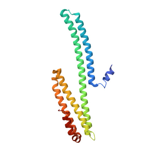
    Beyond the pathology of Alzheimer's disease, the members of the amyloid precursor protein (APP) family are essential for neuronal development and cell homeostasis in mammals. APP and its paralogues APP-like protein 1 (APLP1) and APP-like protein 2 (APLP2) contain the highly conserved heparan sulfate (HS) binding domain E2, which effects various (patho)physiological functions. Here, two crystal structures of the E2 domain of APLP1 are presented in the apo form and in complex with a heparin dodecasaccharide at 2.5 Å resolution. The apo structure of APLP1 E2 revealed an unfolded and hence flexible N-terminal helix αA. The (APLP1 E2)2-(heparin)2 complex structure revealed two distinct binding modes, with APLP1 E2 explicitly recognizing the heparin terminus but also interacting with a continuous heparin chain. The latter only requires a certain register of the sugar moieties that fits to a positively charged surface patch and contributes to the general heparin-binding capability of APP-family proteins. Terminal binding of APLP1 E2 to heparin specifically involves a structure of the nonreducing end that is very similar to heparanase-processed HS chains. These data reveal a conserved mechanism for the binding of APP-family proteins to HS and imply a specific regulatory role of HS modifications in the biology of APP and APP-like proteins.


  • Organizational Affiliation

    Protein Crystallography Group, Leibniz Institute for Age Research (FLI), Beutenbergstrasse 11, 07745 Jena, Germany.


Macromolecules
Find similar proteins by:  (by identity cutoff)  |  3D Structure
Entity ID: 1
MoleculeChains Sequence LengthOrganismDetailsImage
Amyloid-like protein 1
A, B
210Homo sapiensMutation(s): 0 
Gene Names: APLP1
UniProt & NIH Common Fund Data Resources
Find proteins for P51693 (Homo sapiens)
Explore P51693 
Go to UniProtKB:  P51693
PHAROS:  P51693
GTEx:  ENSG00000105290 
Entity Groups  
Sequence Clusters30% Identity50% Identity70% Identity90% Identity95% Identity100% Identity
UniProt GroupP51693
Sequence Annotations
Expand
  • Reference Sequence
Oligosaccharides

Help

Entity ID: 2
MoleculeChains Length2D Diagram Glycosylation3D Interactions
4-deoxy-2-O-sulfo-alpha-L-threo-hex-4-enopyranuronic acid-(1-4)-2-deoxy-6-O-sulfo-2-(sulfoamino)-alpha-D-glucopyranose-(1-4)-2-O-sulfo-alpha-L-idopyranuronic acid-(1-4)-2-deoxy-6-O-sulfo-2-(sulfoamino)-alpha-D-glucopyranose-(1-4)-2-O-sulfo-alpha-L-idopyranuronic acid
C
5N/A
Glycosylation Resources
GlyTouCan:  G25610IB
GlyCosmos:  G25610IB
Entity ID: 3
MoleculeChains Length2D Diagram Glycosylation3D Interactions
2-deoxy-6-O-sulfo-2-(sulfoamino)-alpha-D-glucopyranose-(1-4)-2-O-sulfo-alpha-L-idopyranuronic acid-(1-4)-2-deoxy-6-O-sulfo-2-(sulfoamino)-alpha-D-glucopyranose-(1-4)-2-O-sulfo-alpha-L-idopyranuronic acid-(1-4)-2-deoxy-6-O-sulfo-2-(sulfoamino)-alpha-D-glucopyranose-(1-4)-2-O-sulfo-alpha-L-idopyranuronic acid
D
6N/A
Glycosylation Resources
GlyTouCan:  G82873VJ
GlyCosmos:  G82873VJ
Small Molecules
Ligands 1 Unique
IDChains Name / Formula / InChI Key2D Diagram3D Interactions
ZN
Query on ZN

Download Ideal Coordinates CCD File 
E [auth A]ZINC ION
Zn
PTFCDOFLOPIGGS-UHFFFAOYSA-N
Experimental Data & Validation

Experimental Data

  • Method: X-RAY DIFFRACTION
  • Resolution: 2.50 Å
  • R-Value Free: 0.245 
  • R-Value Work: 0.216 
  • R-Value Observed: 0.218 
  • Space Group: P 31 2 1
Unit Cell:
Length ( Å )Angle ( ˚ )
a = 91.969α = 90
b = 91.969β = 90
c = 208.481γ = 120
Software Package:
Software NamePurpose
MxCuBEdata collection
SHARPphasing
PHENIXrefinement
XDSdata reduction
XDSdata scaling

Structure Validation

View Full Validation Report


Entry History 

Deposition Data

Revision History  (Full details and data files)

  • Version 1.0: 2015-03-11
    Type: Initial release
  • Version 1.1: 2015-03-18
    Changes: Database references
  • Version 2.0: 2020-07-29
    Type: Remediation
    Reason: Carbohydrate remediation
    Changes: Advisory, Atomic model, Data collection, Database references, Derived calculations, Structure summary
  • Version 2.1: 2024-02-28
    Changes: Advisory, Data collection, Database references, Structure summary