4RCH

Discovery of 2-Pyridyl Ureas as Glucokinase Activators


Experimental Data Snapshot

  • Method: X-RAY DIFFRACTION
  • Resolution: 2.30 Å
  • R-Value Free: 0.267 
  • R-Value Work: 0.231 
  • R-Value Observed: 0.233 

wwPDB Validation   3D Report Full Report


Ligand Structure Quality Assessment 


This is version 1.3 of the entry. See complete history


Literature

Discovery of 2-pyridylureas as glucokinase activators.

Hinklin, R.J.Aicher, T.D.Anderson, D.A.Baer, B.R.Boyd, S.A.Condroski, K.R.DeWolf, W.E.Kraser, C.F.McVean, M.Rhodes, S.P.Sturgis, H.L.Voegtli, W.C.Williams, L.Houze, J.B.

(2014) J Med Chem 57: 8180-8186

  • DOI: https://doi.org/10.1021/jm501204z
  • Primary Citation of Related Structures:  
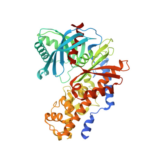
    4RCH

  • PubMed Abstract: 

    Glucokinase (GK) is the rate-limiting step for insulin release from the pancreas in response to high levels of glucose. Flux through GK also contributes to reducing hepatic glucose output. Since many individuals with type 2 diabetes appear to have an inadequacy or defect in one or both of these processes, identifying compounds that can allosterically activate GK may address this issue. Herein we report the identification and initial optimization of a novel series of glucokinase activators (GKAs). Optimization led to the identification of 33 as a compound that displayed activity in an oral glucose tolerance test (OGTT) in normal and diabetic mice.


  • Organizational Affiliation

    Array BioPharma Inc. , 3200 Walnut Street, Boulder, Colorado 80301, United States.


Macromolecules
Find similar proteins by:  (by identity cutoff)  |  3D Structure
Entity ID: 1
MoleculeChains Sequence LengthOrganismDetailsImage
Glucokinase474Homo sapiensMutation(s): 0 
Gene Names: GCK
EC: 2.7.1.2
UniProt & NIH Common Fund Data Resources
Find proteins for P35557 (Homo sapiens)
Explore P35557 
Go to UniProtKB:  P35557
PHAROS:  P35557
GTEx:  ENSG00000106633 
Entity Groups  
Sequence Clusters30% Identity50% Identity70% Identity90% Identity95% Identity100% Identity
UniProt GroupP35557
Sequence Annotations
Expand
  • Reference Sequence
Small Molecules
Ligands 2 Unique
IDChains Name / Formula / InChI Key2D Diagram3D Interactions
3LZ
Query on 3LZ

Download Ideal Coordinates CCD File 
C [auth A]1-{3-[(2-ethylpyridin-3-yl)oxy]-5-(pyridin-2-ylsulfanyl)pyridin-2-yl}-3-methylurea
C19 H19 N5 O2 S
PIGWQTQWPKNWHF-UHFFFAOYSA-N
GLC
Query on GLC

Download Ideal Coordinates CCD File 
B [auth A]alpha-D-glucopyranose
C6 H12 O6
WQZGKKKJIJFFOK-DVKNGEFBSA-N
Binding Affinity Annotations 
IDSourceBinding Affinity
3LZ BindingDB:  4RCH EC50: min: 88, max: 231 (nM) from 2 assay(s)
Experimental Data & Validation

Experimental Data

  • Method: X-RAY DIFFRACTION
  • Resolution: 2.30 Å
  • R-Value Free: 0.267 
  • R-Value Work: 0.231 
  • R-Value Observed: 0.233 
  • Space Group: P 65 2 2
Unit Cell:
Length ( Å )Angle ( ˚ )
a = 80.116α = 90
b = 80.116β = 90
c = 321.807γ = 120
Software Package:
Software NamePurpose
ADSCdata collection
MOLREPphasing
REFMACrefinement
CrystalCleardata reduction
SCALAdata scaling

Structure Validation

View Full Validation Report


Ligand Structure Quality Assessment 


Entry History 

Deposition Data

Revision History  (Full details and data files)

  • Version 1.0: 2014-10-08
    Type: Initial release
  • Version 1.1: 2014-10-22
    Changes: Database references
  • Version 1.2: 2020-07-29
    Type: Remediation
    Reason: Carbohydrate remediation
    Changes: Data collection, Database references, Derived calculations, Structure summary
  • Version 1.3: 2023-09-20
    Changes: Data collection, Database references, Refinement description, Structure summary