4R0M

Structure of McyG A-PCP complexed with phenylalanyl-adenylate


Experimental Data Snapshot

  • Method: X-RAY DIFFRACTION
  • Resolution: 2.45 Å
  • R-Value Free: 0.273 
  • R-Value Work: 0.247 
  • R-Value Observed: 0.248 

wwPDB Validation   3D Report Full Report


Ligand Structure Quality Assessment 


This is version 1.1 of the entry. See complete history


Literature

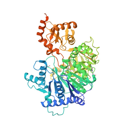
Structure of the adenylation-peptidyl carrier protein didomain of the Microcystis aeruginosa microcystin synthetase McyG.

Tan, X.F.Dai, Y.N.Zhou, K.Jiang, Y.L.Ren, Y.M.Chen, Y.Zhou, C.Z.

(2015) Acta Crystallogr D Biol Crystallogr 71: 873-881

  • DOI: https://doi.org/10.1107/S1399004715001716
  • Primary Citation of Related Structures:  
    4R0M

  • PubMed Abstract: 

    Microcystins, which are the most common cause of hepatotoxicity associated with cyanobacterial water blooms, are assembled in vivo on a large multienzyme complex via a mixed nonribosomal peptide synthetase/polyketide synthetase (NRPS/PKS). The biosynthesis of microcystin in Microcystis aeruginosa PCC 7806 starts with the enzyme McyG, which contains an adenylation-peptidyl carrier protein (A-PCP) didomain for loading the starter unit to assemble the side chain of an Adda residue. However, the catalytic mechanism remains unclear. Here, the 2.45 Å resolution crystal structure of the McyG A-PCP didomain complexed with the catalytic intermediate L-phenylalanyl-adenylate (L-Phe-AMP) is reported. Each asymmetric unit contains two protein molecules, one of which consists of the A-PCP didomain and the other of which comprises only the A domain. Structural analyses suggest that Val227 is likely to be critical for the selection of hydrophobic substrates. Moreover, two distinct interfaces demonstrating variable crosstalk between the PCP domain and the A domain were observed. A catalytic cycle for the adenylation and peptide transfer of the A-PCP didomain is proposed.


  • Organizational Affiliation

    Hefei National Laboratory for Physical Sciences at the Microscale and School of Life Sciences, University of Science and Technology of China, Hefei, Anhui 230027, People's Republic of China.


Macromolecules
Find similar proteins by:  (by identity cutoff)  |  3D Structure
Entity ID: 1
MoleculeChains Sequence LengthOrganismDetailsImage
McyG proteinA [auth B],
B [auth A]
643Microcystis aeruginosa PCC 7806Mutation(s): 0 
Gene Names: mcyGIPF_370
UniProt
Find proteins for A8YJW1 (Microcystis aeruginosa (strain PCC 7806))
Explore A8YJW1 
Go to UniProtKB:  A8YJW1
Entity Groups  
Sequence Clusters30% Identity50% Identity70% Identity90% Identity95% Identity100% Identity
UniProt GroupA8YJW1
Sequence Annotations
Expand
  • Reference Sequence
Small Molecules
Ligands 1 Unique
IDChains Name / Formula / InChI Key2D Diagram3D Interactions
FA5
Query on FA5

Download Ideal Coordinates CCD File 
C [auth B],
D [auth A]
ADENOSINE-5'-[PHENYLALANINYL-PHOSPHATE]
C19 H23 N6 O8 P
ZFNOOEXYTZPIQS-URQYDQELSA-N
Modified Residues  1 Unique
IDChains TypeFormula2D DiagramParent
MSE
Query on MSE
A [auth B],
B [auth A]
L-PEPTIDE LINKINGC5 H11 N O2 SeMET
Experimental Data & Validation

Experimental Data

  • Method: X-RAY DIFFRACTION
  • Resolution: 2.45 Å
  • R-Value Free: 0.273 
  • R-Value Work: 0.247 
  • R-Value Observed: 0.248 
  • Space Group: P 32 2 1
Unit Cell:
Length ( Å )Angle ( ˚ )
a = 120.71α = 90
b = 120.71β = 90
c = 262.78γ = 120
Software Package:
Software NamePurpose
ADSCdata collection
SOLVEphasing
REFMACrefinement
MOSFLMdata reduction
SCALAdata scaling

Structure Validation

View Full Validation Report


Ligand Structure Quality Assessment 


Entry History 

Deposition Data

Revision History  (Full details and data files)

  • Version 1.0: 2015-04-15
    Type: Initial release
  • Version 1.1: 2019-12-18
    Changes: Database references, Derived calculations