4QYS

TrpB2 enzymes


Experimental Data Snapshot

  • Method: X-RAY DIFFRACTION
  • Resolution: 1.94 Å
  • R-Value Free: 0.252 
  • R-Value Work: 0.195 
  • R-Value Observed: 0.198 

wwPDB Validation   3D Report Full Report


Ligand Structure Quality Assessment 


This is version 1.1 of the entry. See complete history


Literature

TrpB2 enzymes are O-phospho-l-serine dependent tryptophan synthases

Busch, F.Rajendran, C.Mayans, O.Loffler, P.Merkl, R.Sterner, R.

(2014) Biochemistry 53: 6078-6083

  • DOI: https://doi.org/10.1021/bi500977y
  • Primary Citation of Related Structures:  
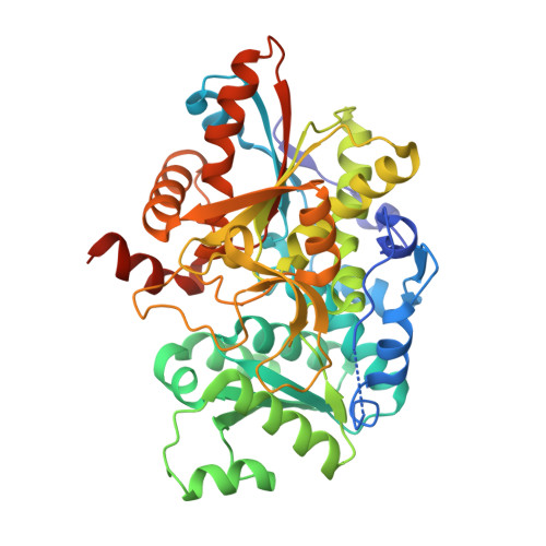
    4QYS

  • PubMed Abstract: 

    The rapid increase of the number of sequenced genomes asks for the functional annotation of the encoded enzymes. We used a combined computational-structural approach to determine the function of the TrpB2 subgroup of the tryptophan synthase β chain/β chain-like TrpB1-TrpB2 family (IPR023026). The results showed that TrpB2 enzymes are O-phospho-l-serine dependent tryptophan synthases, whereas TrpB1 enzymes catalyze the l-serine dependent synthesis of tryptophan. We found a single residue being responsible for the different substrate specificities of TrpB1 and TrpB2 and confirmed this finding by mutagenesis studies and crystallographic analysis of a TrpB2 enzyme with bound O-phospho-l-serine.


  • Organizational Affiliation

    Institute of Biophysics and Physical Biochemistry, University of Regensburg , Universitätsstrasse 31, D-93053 Regensburg, Germany.


Macromolecules
Find similar proteins by:  (by identity cutoff)  |  3D Structure
Entity ID: 1
MoleculeChains Sequence LengthOrganismDetailsImage
Tryptophan synthase beta chain 2A [auth B],
B [auth A]
442Saccharolobus solfataricus P2Mutation(s): 0 
Gene Names: trpB2SSO1145
EC: 4.2.1.20
UniProt
Find proteins for Q97TX6 (Saccharolobus solfataricus (strain ATCC 35092 / DSM 1617 / JCM 11322 / P2))
Explore Q97TX6 
Go to UniProtKB:  Q97TX6
Entity Groups  
Sequence Clusters30% Identity50% Identity70% Identity90% Identity95% Identity100% Identity
UniProt GroupQ97TX6
Sequence Annotations
Expand
  • Reference Sequence
Small Molecules
Ligands 3 Unique
IDChains Name / Formula / InChI Key2D Diagram3D Interactions
PLP
Query on PLP

Download Ideal Coordinates CCD File 
D [auth A]PYRIDOXAL-5'-PHOSPHATE
C8 H10 N O6 P
NGVDGCNFYWLIFO-UHFFFAOYSA-N
PLR
Query on PLR

Download Ideal Coordinates CCD File 
C [auth B](5-HYDROXY-4,6-DIMETHYLPYRIDIN-3-YL)METHYL DIHYDROGEN PHOSPHATE
C8 H12 N O5 P
RBCOYOYDYNXAFA-UHFFFAOYSA-N
SEP
Query on SEP

Download Ideal Coordinates CCD File 
E [auth A]PHOSPHOSERINE
C3 H8 N O6 P
BZQFBWGGLXLEPQ-REOHCLBHSA-N
Experimental Data & Validation

Experimental Data

  • Method: X-RAY DIFFRACTION
  • Resolution: 1.94 Å
  • R-Value Free: 0.252 
  • R-Value Work: 0.195 
  • R-Value Observed: 0.198 
  • Space Group: P 1 21 1
Unit Cell:
Length ( Å )Angle ( ˚ )
a = 55.564α = 90
b = 61.903β = 98.23
c = 109.574γ = 90
Software Package:
Software NamePurpose
PHENIXrefinement

Structure Validation

View Full Validation Report


Ligand Structure Quality Assessment 


Entry History 

Deposition Data

Revision History  (Full details and data files)

  • Version 1.0: 2015-02-18
    Type: Initial release
  • Version 1.1: 2024-03-20
    Changes: Data collection, Database references, Derived calculations