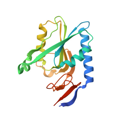
1.95 Angstrom resolution crystal structure of a hypoxanthine-guanine phosphoribosyltransferase (hpt-2) from Bacillus anthracis str. 'Ames Ancestor' with HEPES molecule in the active site
Halavaty, A.S., Minasov, G., Dubrovska, I., Winsor, J., Shuvalova, L., Anderson, W.F., Center for Structural Genomics of Infectious Diseases (CSGID)To be published.