4PYQ

Humanized rat apo-COMT in complex with a ureido-benzamidine


Experimental Data Snapshot

  • Method: X-RAY DIFFRACTION
  • Resolution: 1.39 Å
  • R-Value Free: 0.161 
  • R-Value Work: 0.132 
  • R-Value Observed: 0.143 

wwPDB Validation   3D Report Full Report


Ligand Structure Quality Assessment 


This is version 1.2 of the entry. See complete history


Literature

Mapping the conformational space accessible to catechol-O-methyltransferase.

Ehler, A.Benz, J.Schlatter, D.Rudolph, M.G.

(2014) Acta Crystallogr D Biol Crystallogr 70: 2163-2174

  • DOI: https://doi.org/10.1107/S1399004714012917
  • Primary Citation of Related Structures:  
    4P7F, 4P7G, 4P7J, 4P7K, 4PYI, 4PYJ, 4PYK, 4PYL, 4PYM, 4PYN, 4PYO, 4PYQ

  • PubMed Abstract: 

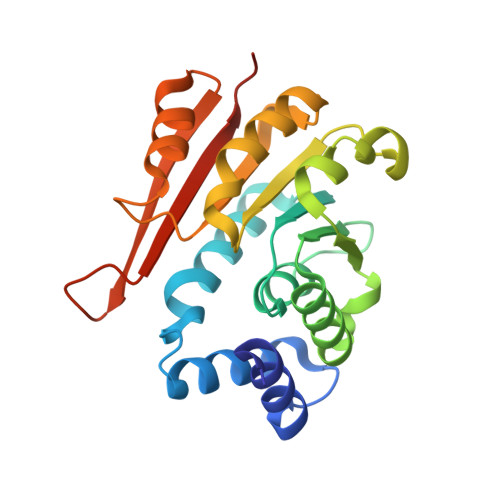
    Methylation catalysed by catechol-O-methyltransferase (COMT) is the main pathway of catechol neurotransmitter deactivation in the prefrontal cortex. Low levels of this class of neurotransmitters are held to be causative of diseases such as schizophrenia, depression and Parkinson's disease. Inhibition of COMT may increase neurotransmitter levels, thus offering a route for treatment. Structure-based drug design hitherto seems to be based on the closed enzyme conformation. Here, a set of apo, semi-holo, holo and Michaelis form crystal structures are described that define the conformational space available to COMT and that include likely intermediates along the catalytic pathway. Domain swaps and sizeable loop movements around the active site testify to the flexibility of this enzyme, rendering COMT a difficult drug target. The low affinity of the co-substrate S-adenosylmethionine and the large conformational changes involved during catalysis highlight significant energetic investment to achieve the closed conformation. Since each conformation of COMT is a bona fide target for inhibitors, other states than the closed conformation may be promising to address. Crystallographic data for an alternative avenue of COMT inhibition, i.e. locking of the apo state by an inhibitor, are presented. The set of COMT structures may prove to be useful for the development of novel classes of inhibitors.


  • Organizational Affiliation

    Molecular Design and Chemical Biology, F. Hoffmann-La Roche, Grenzacher Strasse 124, Basel, Switzerland.


Macromolecules
Find similar proteins by:  (by identity cutoff)  |  3D Structure
Entity ID: 1
MoleculeChains Sequence LengthOrganismDetailsImage
Catechol O-methyltransferase
A, B
221Rattus norvegicusMutation(s): 2 
Gene Names: Comt
EC: 2.1.1.6
UniProt
Find proteins for P22734 (Rattus norvegicus)
Explore P22734 
Go to UniProtKB:  P22734
Entity Groups  
Sequence Clusters30% Identity50% Identity70% Identity90% Identity95% Identity100% Identity
UniProt GroupP22734
Sequence Annotations
Expand
  • Reference Sequence
Small Molecules
Ligands 5 Unique
IDChains Name / Formula / InChI Key2D Diagram3D Interactions
2X1
Query on 2X1

Download Ideal Coordinates CCD File 
C [auth A],
I [auth B]
4-({[3-(aminomethyl)phenyl]carbamoyl}amino)benzenecarboximidamide
C15 H17 N5 O
LNSAAROLFORION-UHFFFAOYSA-N
SO4
Query on SO4

Download Ideal Coordinates CCD File 
F [auth A],
G [auth A],
L [auth B],
M [auth B]
SULFATE ION
O4 S
QAOWNCQODCNURD-UHFFFAOYSA-L
ACT
Query on ACT

Download Ideal Coordinates CCD File 
D [auth A],
J [auth B]
ACETATE ION
C2 H3 O2
QTBSBXVTEAMEQO-UHFFFAOYSA-M
CL
Query on CL

Download Ideal Coordinates CCD File 
K [auth B]CHLORIDE ION
Cl
VEXZGXHMUGYJMC-UHFFFAOYSA-M
NA
Query on NA

Download Ideal Coordinates CCD File 
E [auth A],
H [auth B]
SODIUM ION
Na
FKNQFGJONOIPTF-UHFFFAOYSA-N
Binding Affinity Annotations 
IDSourceBinding Affinity
2X1 Binding MOAD:  4PYQ IC50: 4400 (nM) from 1 assay(s)
Experimental Data & Validation

Experimental Data

  • Method: X-RAY DIFFRACTION
  • Resolution: 1.39 Å
  • R-Value Free: 0.161 
  • R-Value Work: 0.132 
  • R-Value Observed: 0.143 
  • Space Group: H 3
Unit Cell:
Length ( Å )Angle ( ˚ )
a = 125.366α = 90
b = 125.366β = 90
c = 77.287γ = 120
Software Package:
Software NamePurpose
PHASERphasing
PHENIXrefinement
XDSdata reduction
SADABSdata scaling

Structure Validation

View Full Validation Report


Ligand Structure Quality Assessment 


Entry History 

Deposition Data

Revision History  (Full details and data files)

  • Version 1.0: 2014-06-11
    Type: Initial release
  • Version 1.1: 2014-08-27
    Changes: Database references
  • Version 1.2: 2024-02-28
    Changes: Data collection, Database references, Derived calculations