4PNE

Crystal Structure of the [4+2]-Cyclase SpnF


Experimental Data Snapshot

  • Method: X-RAY DIFFRACTION
  • Resolution: 1.50 Å
  • R-Value Free: 0.220 
  • R-Value Work: 0.197 
  • R-Value Observed: 0.198 

wwPDB Validation   3D Report Full Report


Ligand Structure Quality Assessment 


This is version 1.6 of the entry. See complete history


Literature

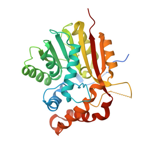
The structure of SpnF, a standalone enzyme that catalyzes [4 + 2] cycloaddition.

Fage, C.D.Isiorho, E.A.Liu, Y.Wagner, D.T.Liu, H.W.Keatinge-Clay, A.T.

(2015) Nat Chem Biol 11: 256-258

  • DOI: https://doi.org/10.1038/nchembio.1768
  • Primary Citation of Related Structures:  
    4PNE

  • PubMed Abstract: 

    In the biosynthetic pathway of the spinosyn insecticides, the tailoring enzyme SpnF performs a [4 + 2] cycloaddition on a 22-membered macrolactone to forge an embedded cyclohexene ring. To learn more about this reaction, which could potentially proceed through a Diels-Alder mechanism, we determined the 1.50-Å-resolution crystal structure of SpnF bound to S-adenosylhomocysteine. This sets the stage for advanced experimental and computational studies to determine the precise mechanism of SpnF-mediated cyclization.


  • Organizational Affiliation

    Department of Molecular Biosciences, The University of Texas at Austin, Austin, Texas, USA.


Macromolecules
Find similar proteins by:  (by identity cutoff)  |  3D Structure
Entity ID: 1
MoleculeChains Sequence LengthOrganismDetailsImage
Methyltransferase-like protein
A, B
302Saccharopolyspora spinosaMutation(s): 0 
Gene Names: spnF
UniProt
Find proteins for Q9ALM7 (Saccharopolyspora spinosa)
Explore Q9ALM7 
Go to UniProtKB:  Q9ALM7
Entity Groups  
Sequence Clusters30% Identity50% Identity70% Identity90% Identity95% Identity100% Identity
UniProt GroupQ9ALM7
Sequence Annotations
Expand
  • Reference Sequence
Experimental Data & Validation

Experimental Data

  • Method: X-RAY DIFFRACTION
  • Resolution: 1.50 Å
  • R-Value Free: 0.220 
  • R-Value Work: 0.197 
  • R-Value Observed: 0.198 
  • Space Group: P 1 21 1
Unit Cell:
Length ( Å )Angle ( ˚ )
a = 41.74α = 90
b = 69.35β = 97.49
c = 83.63γ = 90
Software Package:
Software NamePurpose
PHENIXrefinement
XDSdata scaling
XSCALEdata scaling
PDB_EXTRACTdata extraction
PHASERphasing

Structure Validation

View Full Validation Report


Ligand Structure Quality Assessment 


Entry History & Funding Information

Deposition Data


Funding OrganizationLocationGrant Number
National Institutes of Health/National Institute of General Medical Sciences (NIH/NIGMS)United StatesGM106112
National Institutes of Health/National Institute of General Medical Sciences (NIH/NIGMS)United StatesGM35906
Welch FoundationUnited StatesF-1712
Welch FoundationUnited StatesF-1511

Revision History  (Full details and data files)

  • Version 1.0: 2015-02-18
    Type: Initial release
  • Version 1.1: 2015-03-11
    Changes: Database references
  • Version 1.2: 2015-04-01
    Changes: Database references
  • Version 1.3: 2017-09-13
    Changes: Author supporting evidence, Derived calculations, Other, Source and taxonomy
  • Version 1.4: 2017-11-22
    Changes: Refinement description
  • Version 1.5: 2019-12-25
    Changes: Author supporting evidence
  • Version 1.6: 2023-09-27
    Changes: Data collection, Database references, Refinement description