4PFJ

The structure of bi-acetylated SAHH


Experimental Data Snapshot

  • Method: X-RAY DIFFRACTION
  • Resolution: 2.30 Å
  • R-Value Free: 0.245 
  • R-Value Work: 0.194 
  • R-Value Observed: 0.196 

wwPDB Validation   3D Report Full Report


Ligand Structure Quality Assessment 


This is version 3.0 of the entry. See complete history


Literature

Regulation of s-adenosylhomocysteine hydrolase by lysine acetylation.

Wang, Y.Kavran, J.M.Chen, Z.Karukurichi, K.R.Leahy, D.J.Cole, P.A.

(2014) J Biol Chem 289: 31361-31372

  • DOI: https://doi.org/10.1074/jbc.M114.597153
  • Primary Citation of Related Structures:  
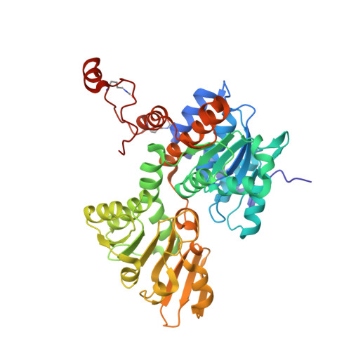
    4PFJ, 4PGF

  • PubMed Abstract: 

    S-Adenosylhomocysteine hydrolase (SAHH) is an NAD(+)-dependent tetrameric enzyme that catalyzes the breakdown of S-adenosylhomocysteine to adenosine and homocysteine and is important in cell growth and the regulation of gene expression. Loss of SAHH function can result in global inhibition of cellular methyltransferase enzymes because of high levels of S-adenosylhomocysteine. Prior proteomics studies have identified two SAHH acetylation sites at Lys(401) and Lys(408) but the impact of these post-translational modifications has not yet been determined. Here we use expressed protein ligation to produce semisynthetic SAHH acetylated at Lys(401) and Lys(408) and show that modification of either position negatively impacts the catalytic activity of SAHH. X-ray crystal structures of 408-acetylated SAHH and dually acetylated SAHH have been determined and reveal perturbations in the C-terminal hydrogen bonding patterns, a region of the protein important for NAD(+) binding. These crystal structures along with mutagenesis data suggest that such hydrogen bond perturbations are responsible for SAHH catalytic inhibition by acetylation. These results suggest how increased acetylation of SAHH may globally influence cellular methylation patterns.


  • Organizational Affiliation

    From the Deptartments of Pharmacology and Molecular Sciences and.


Macromolecules
Find similar proteins by:  (by identity cutoff)  |  3D Structure
Entity ID: 1
MoleculeChains Sequence LengthOrganismDetailsImage
Adenosylhomocysteinase
A, B
432Homo sapiensMutation(s): 2 
Gene Names: AHCYSAHH
EC: 3.3.1.1
UniProt & NIH Common Fund Data Resources
Find proteins for P23526 (Homo sapiens)
Explore P23526 
Go to UniProtKB:  P23526
PHAROS:  P23526
GTEx:  ENSG00000101444 
Entity Groups  
Sequence Clusters30% Identity50% Identity70% Identity90% Identity95% Identity100% Identity
UniProt GroupP23526
Sequence Annotations
Expand
  • Reference Sequence
Small Molecules
Experimental Data & Validation

Experimental Data

  • Method: X-RAY DIFFRACTION
  • Resolution: 2.30 Å
  • R-Value Free: 0.245 
  • R-Value Work: 0.194 
  • R-Value Observed: 0.196 
  • Space Group: I 2 2 2
Unit Cell:
Length ( Å )Angle ( ˚ )
a = 98.366α = 90
b = 102.727β = 90
c = 176.16γ = 90
Software Package:
Software NamePurpose
PHENIXrefinement
PHASERphasing

Structure Validation

View Full Validation Report


Ligand Structure Quality Assessment 


Entry History 

Deposition Data

Revision History  (Full details and data files)

  • Version 1.0: 2014-10-01
    Type: Initial release
  • Version 1.1: 2014-10-08
    Changes: Database references
  • Version 1.2: 2014-11-19
    Changes: Database references
  • Version 1.3: 2017-11-22
    Changes: Advisory, Database references, Derived calculations, Other, Refinement description, Source and taxonomy
  • Version 2.0: 2018-01-17
    Changes: Advisory, Atomic model, Data collection, Derived calculations, Refinement description
  • Version 2.1: 2023-09-27
    Changes: Advisory, Data collection, Database references, Refinement description
  • Version 3.0: 2023-11-15
    Changes: Atomic model, Data collection