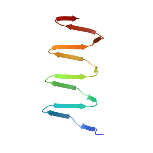
4OSD

Dimer of a C-terminal fragment of phage T4 gp5 beta-helix


Experimental Data Snapshot

  • Method: X-RAY DIFFRACTION
  • Resolution: 1.96 Å
  • R-Value Free: 0.279 
  • R-Value Work: 0.219 
  • R-Value Observed: 0.222 

wwPDB Validation   3D Report Full Report


Ligand Structure Quality Assessment 


This is version 1.3 of the entry. See complete history


Literature

Structure and Biophysical Properties of a Triple-Stranded Beta-Helix Comprising the Central Spike of Bacteriophage T4.

Buth, S.A.Menin, L.Shneider, M.M.Engel, J.Boudko, S.P.Leiman, P.G.

(2015) Viruses 7: 4676-4706

  • DOI: https://doi.org/10.3390/v7082839
  • Primary Citation of Related Structures:  
    4JJ2, 4OSD

  • PubMed Abstract: 

    Gene product 5 (gp5) of bacteriophage T4 is a spike-shaped protein that functions to disrupt the membrane of the target cell during phage infection. Its C-terminal domain is a long and slender β-helix that is formed by three polypeptide chains wrapped around a common symmetry axis akin to three interdigitated corkscrews. The folding and biophysical properties of such triple-stranded β-helices, which are topologically related to amyloid fibers, represent an unsolved biophysical problem. Here, we report structural and biophysical characterization of T4 gp5 β-helix and its truncated mutants of different lengths. A soluble fragment that forms a dimer of trimers and that could comprise a minimal self-folding unit has been identified. Surprisingly, the hydrophobic core of the β-helix is small. It is located near the C-terminal end of the β-helix and contains a centrally positioned and hydrated magnesium ion. A large part of the β-helix interior comprises a large elongated cavity that binds palmitic, stearic, and oleic acids in an extended conformation suggesting that these molecules might participate in the folding of the complete β-helix.


  • Organizational Affiliation

    Institute of Physics of Biological Systems, École Polytechnique Fédérale de Lausanne (EPFL), BSP 415, 1015 Lausanne, Switzerland. sergii.buth@epfl.ch.


Macromolecules
Find similar proteins by:  (by identity cutoff)  |  3D Structure
Entity ID: 1
MoleculeChains Sequence LengthOrganismDetailsImage
Tail-associated lysozyme
A, B, C, D, E
A, B, C, D, E, F, G, H, I, J, K, L, M, N, O, P, Q, R
95Tequatrovirus T4Mutation(s): 0 
Gene Names: 5
EC: 3.2.1.17
UniProt
Find proteins for P16009 (Enterobacteria phage T4)
Explore P16009 
Go to UniProtKB:  P16009
Entity Groups  
Sequence Clusters30% Identity50% Identity70% Identity90% Identity95% Identity100% Identity
UniProt GroupP16009
Sequence Annotations
Expand
  • Reference Sequence
Small Molecules
Ligands 4 Unique
IDChains Name / Formula / InChI Key2D Diagram3D Interactions
STE
Query on STE

Download Ideal Coordinates CCD File 
AA [auth G]
FA [auth K]
LA [auth O]
MA [auth P]
V [auth C]
AA [auth G],
FA [auth K],
LA [auth O],
MA [auth P],
V [auth C],
Y [auth F]
STEARIC ACID
C18 H36 O2
QIQXTHQIDYTFRH-UHFFFAOYSA-N
ELA
Query on ELA

Download Ideal Coordinates CCD File 
BA [auth G]
EA [auth J]
IA [auth M]
NA [auth P]
S [auth A]
BA [auth G],
EA [auth J],
IA [auth M],
NA [auth P],
S [auth A],
T [auth A],
Z [auth F]
9-OCTADECENOIC ACID
C18 H34 O2
ZQPPMHVWECSIRJ-MDZDMXLPSA-N
PLM
Query on PLM

Download Ideal Coordinates CCD File 
DA [auth H],
GA [auth K],
JA [auth N],
PA [auth Q],
W [auth D]
PALMITIC ACID
C16 H32 O2
IPCSVZSSVZVIGE-UHFFFAOYSA-N
MG
Query on MG

Download Ideal Coordinates CCD File 
CA [auth H]
HA [auth L]
KA [auth O]
OA [auth Q]
U [auth B]
CA [auth H],
HA [auth L],
KA [auth O],
OA [auth Q],
U [auth B],
X [auth F]
MAGNESIUM ION
Mg
JLVVSXFLKOJNIY-UHFFFAOYSA-N
Experimental Data & Validation

Experimental Data

  • Method: X-RAY DIFFRACTION
  • Resolution: 1.96 Å
  • R-Value Free: 0.279 
  • R-Value Work: 0.219 
  • R-Value Observed: 0.222 
  • Space Group: P 1 21 1
Unit Cell:
Length ( Å )Angle ( ˚ )
a = 110.288α = 90
b = 73.786β = 113.39
c = 111.602γ = 90
Software Package:
Software NamePurpose
PHASERphasing
REFMACrefinement
XDSdata reduction
XDSdata scaling

Structure Validation

View Full Validation Report


Ligand Structure Quality Assessment 


Entry History 

Deposition Data

Revision History  (Full details and data files)

  • Version 1.0: 2015-02-18
    Type: Initial release
  • Version 1.1: 2017-11-22
    Changes: Refinement description
  • Version 1.2: 2023-02-08
    Changes: Database references, Derived calculations
  • Version 1.3: 2023-11-08
    Changes: Data collection, Refinement description