4OMJ

Crystal structure of SPF bound to 2,3-oxidosqualene


Experimental Data Snapshot

  • Method: X-RAY DIFFRACTION
  • Resolution: 1.60 Å
  • R-Value Free: 0.179 
  • R-Value Work: 0.154 
  • R-Value Observed: 0.156 

wwPDB Validation   3D Report Full Report


Ligand Structure Quality Assessment 


This is version 1.2 of the entry. See complete history


Literature

Structural insights on cholesterol endosynthesis: Binding of squalene and 2,3-oxidosqualene to supernatant protein factor.

Christen, M.Marcaida, M.J.Lamprakis, C.Aeschimann, W.Vaithilingam, J.Schneider, P.Hilbert, M.Schneider, G.Cascella, M.Stocker, A.

(2015) J Struct Biol 190: 261-270

  • DOI: https://doi.org/10.1016/j.jsb.2015.05.001
  • Primary Citation of Related Structures:  
    4OMJ, 4OMK

  • PubMed Abstract: 

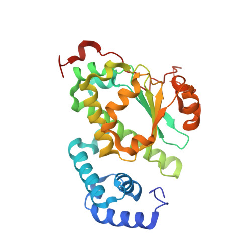
    We present the crystal structures of the SEC14-like domain of supernatant protein factor (SPF) in complex with squalene and 2,3-oxidosqualene. The structures were resolved at 1.75Å (complex with squalene) and 1.6Å resolution (complex with 2,3-oxidosqualene), leading in both cases to clear images of the protein/substrate interactions. Ligand binding is facilitated by removal of the Golgi-dynamics (GOLD) C-terminal domain of SPF, which, as shown in previous structures of the apo-protein, blocked the opening of the binding pocket to the exterior. Both substrates bind into a large hydrophobic cavity, typical of such lipid-transporter family. Our structures report no specific recognition mode for the epoxide group. In fact, for both molecules, ligand affinity is dominated by hydrophobic interactions, and independent investigations by computational models or differential scanning micro-calorimetry reveal similar binding affinities for both ligands. Our findings elucidate the molecular bases of the role of SPF in sterol endo-synthesis, supporting the original hypothesis that SPF is a facilitator of substrate flow within the sterol synthetic pathway. Moreover, our results suggest that the GOLD domain acts as a regulator, as its conformational displacement must occur to favor ligand binding and release during the different synthetic steps.


  • Organizational Affiliation

    Department of Chemistry and Biochemistry, University of Bern, Freiestrasse 3, CH-3012 Bern, Switzerland.


Macromolecules
Find similar proteins by:  (by identity cutoff)  |  3D Structure
Entity ID: 1
MoleculeChains Sequence LengthOrganismDetailsImage
SEC14-like protein 2
A, B
278Homo sapiensMutation(s): 0 
Gene Names: C22orf6KIAA1186KIAA1658SEC14L2
UniProt & NIH Common Fund Data Resources
Find proteins for O76054 (Homo sapiens)
Explore O76054 
Go to UniProtKB:  O76054
PHAROS:  O76054
GTEx:  ENSG00000100003 
Entity Groups  
Sequence Clusters30% Identity50% Identity70% Identity90% Identity95% Identity100% Identity
UniProt GroupO76054
Sequence Annotations
Expand
  • Reference Sequence
Small Molecules
Ligands 3 Unique
IDChains Name / Formula / InChI Key2D Diagram3D Interactions
2TX
Query on 2TX

Download Ideal Coordinates CCD File 
L [auth A],
T [auth B]
(3S)-2,2-dimethyl-3-[(3E,7E,11E,15E)-3,7,12,16,20-pentamethylhenicosa-3,7,11,15,19-pentaen-1-yl]oxirane
C30 H50 O
QYIMSPSDBYKPPY-RSKUXYSASA-N
SO4
Query on SO4

Download Ideal Coordinates CCD File 
C [auth A]
D [auth A]
E [auth A]
M [auth B]
N [auth B]
C [auth A],
D [auth A],
E [auth A],
M [auth B],
N [auth B],
O [auth B],
P [auth B]
SULFATE ION
O4 S
QAOWNCQODCNURD-UHFFFAOYSA-L
CL
Query on CL

Download Ideal Coordinates CCD File 
F [auth A]
G [auth A]
H [auth A]
I [auth A]
J [auth A]
F [auth A],
G [auth A],
H [auth A],
I [auth A],
J [auth A],
K [auth A],
Q [auth B],
R [auth B],
S [auth B]
CHLORIDE ION
Cl
VEXZGXHMUGYJMC-UHFFFAOYSA-M
Binding Affinity Annotations 
IDSourceBinding Affinity
2TX Binding MOAD:  4OMJ Kd: 5440 (nM) from 1 assay(s)
Experimental Data & Validation

Experimental Data

  • Method: X-RAY DIFFRACTION
  • Resolution: 1.60 Å
  • R-Value Free: 0.179 
  • R-Value Work: 0.154 
  • R-Value Observed: 0.156 
  • Space Group: P 1 21 1
Unit Cell:
Length ( Å )Angle ( ˚ )
a = 71.88α = 90
b = 73.8β = 96.29
c = 94.45γ = 90
Software Package:
Software NamePurpose
XDSdata scaling
PHASERphasing
PHENIXrefinement
MOSFLMdata reduction
SCALAdata scaling

Structure Validation

View Full Validation Report


Ligand Structure Quality Assessment 


Entry History 

Deposition Data

Revision History  (Full details and data files)

  • Version 1.0: 2015-04-15
    Type: Initial release
  • Version 1.1: 2015-06-24
    Changes: Database references
  • Version 1.2: 2023-09-20
    Changes: Data collection, Database references, Derived calculations, Refinement description