Crystal Structure of Calcium Binding Protein-5 from Entamoeba histolytica and Its Involvement in Initiation of Phagocytosis of Human Erythrocytes.
Kumar, S., Aslam, S., Mazumder, M., Dahiya, P., Murmu, A., Manjasetty, B.A., Zaidi, R., Bhattacharya, A., Gourinath, S.(2014) PLoS Pathog 10: e1004532-e1004532
- PubMed: 25502654
- DOI: https://doi.org/10.1371/journal.ppat.1004532
- Primary Citation of Related Structures:
4OCI - PubMed Abstract:
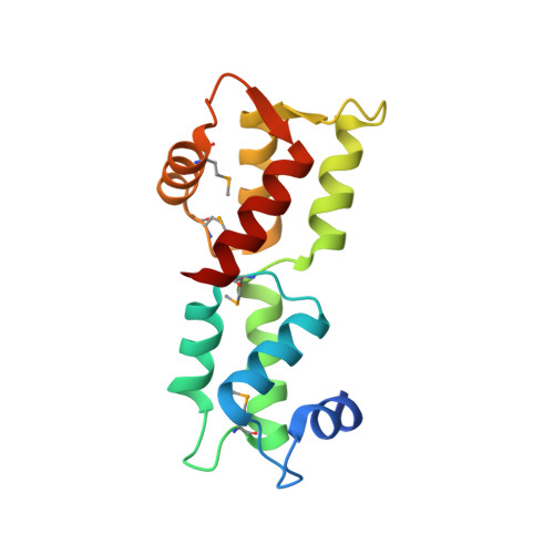
Entamoeba histolytica is the etiological agent of human amoebic colitis and liver abscess, and causes a high level of morbidity and mortality worldwide, particularly in developing countries. There are a number of studies that have shown a crucial role for Ca2+ and its binding protein in amoebic biology. EhCaBP5 is one of the EF hand calcium-binding proteins of E. histolytica. We have determined the crystal structure of EhCaBP5 at 1.9 Å resolution in the Ca2+-bound state, which shows an unconventional mode of Ca2+ binding involving coordination to a closed yet canonical EF-hand motif. Structurally, EhCaBP5 is more similar to the essential light chain of myosin than to Calmodulin despite its somewhat greater sequence identity with Calmodulin. This structure-based analysis suggests that EhCaBP5 could be a light chain of myosin. Surface plasmon resonance studies confirmed this hypothesis, and in particular showed that EhCaBP5 interacts with the IQ motif of myosin 1B in calcium independent manner. It also appears from modelling of the EhCaBP5-IQ motif complex that EhCaBP5 undergoes a structural change in order to bind the IQ motif of myosin. This specific interaction was further confirmed by the observation that EhCaBP5 and myosin 1B are colocalized in E. histolytica during phagocytic cup formation. Immunoprecipitation of EhCaBP5 from total E. histolytica cellular extract also pulls out myosin 1B and this interaction was confirmed to be Ca2+ independent. Confocal imaging of E. histolytica showed that EhCaBP5 and myosin 1B are part of phagosomes. Overexpression of EhCaBP5 increases slight rate (∼20%) of phagosome formation, while suppression reduces the rate drastically (∼55%). Taken together, these experiments indicate that EhCaBP5 is likely to be the light chain of myosin 1B. Interestingly, EhCaBP5 is not present in the phagosome after its formation suggesting EhCaBP5 may be playing a regulatory role.
Organizational Affiliation:
School of Life Sciences, Jawaharlal Nehru University, New Delhi, India; Department of Biochemistry, Jamia Hamdard, New Delhi, India.