4O0L

Crystal structure of NADPH-Dependent 3-Quinuclidinone Reductase from Rhodotorula Rubra


Experimental Data Snapshot

  • Method: X-RAY DIFFRACTION
  • Resolution: 2.20 Å
  • R-Value Free: 0.223 
  • R-Value Work: 0.182 
  • R-Value Observed: 0.184 

wwPDB Validation   3D Report Full Report


Ligand Structure Quality Assessment 


This is version 1.1 of the entry. See complete history


Literature

Structural basis of stereospecific reduction by quinuclidinone reductase

Takeshita, D.Kataoka, M.Miyakawa, T.Miyazono, K.Kumashiro, S.Nagai, T.Urano, N.Uzura, A.Nagata, K.Shimizu, S.Tanokura, M.

(2014) AMB Express 4: 6-6

  • DOI: https://doi.org/10.1186/2191-0855-4-6
  • Primary Citation of Related Structures:  
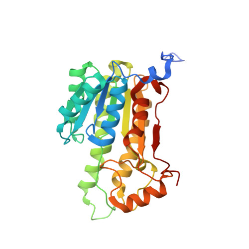
    4O0L

  • PubMed Abstract: 

    Chiral molecule (R)-3-quinuclidinol, a valuable compound for the production of various pharmaceuticals, is efficiently synthesized from 3-quinuclidinone by using NADPH-dependent 3-quinuclidinone reductase (RrQR) from Rhodotorula rubra. Here, we report the crystal structure of RrQR and the structure-based mutational analysis. The enzyme forms a tetramer, in which the core of each protomer exhibits the α/β Rossmann fold and contains one molecule of NADPH, whereas the characteristic substructures of a small lobe and a variable loop are localized around the substrate-binding site. Modeling and mutation analyses of the catalytic site indicated that the hydrophobicity of two residues, I167 and F212, determines the substrate-binding orientation as well as the substrate-binding affinity. Our results revealed that the characteristic substrate-binding pocket composed of hydrophobic amino acid residues ensures substrate docking for the stereospecific reaction of RrQR in spite of its loose interaction with the substrate.


  • Organizational Affiliation

    Department of Applied Biological Chemistry, Graduate School of Agricultural and Life Sciences, University of Tokyo, 1-1-1 Yayoi, Bunkyo-ku, Tokyo 113-8657, Japan. amtanok@mail.ecc.u-tokyo.ac.jp.


Macromolecules
Find similar proteins by:  (by identity cutoff)  |  3D Structure
Entity ID: 1
MoleculeChains Sequence LengthOrganismDetailsImage
NADPH-dependent 3-quinuclidinone reductase
A, B, C, D
275Rhodotorula mucilaginosaMutation(s): 0 
Gene Names: rqr
UniProt
Find proteins for B9ZZZ6 (Rhodotorula mucilaginosa)
Explore B9ZZZ6 
Go to UniProtKB:  B9ZZZ6
Entity Groups  
Sequence Clusters30% Identity50% Identity70% Identity90% Identity95% Identity100% Identity
UniProt GroupB9ZZZ6
Sequence Annotations
Expand
  • Reference Sequence
Experimental Data & Validation

Experimental Data

  • Method: X-RAY DIFFRACTION
  • Resolution: 2.20 Å
  • R-Value Free: 0.223 
  • R-Value Work: 0.182 
  • R-Value Observed: 0.184 
  • Space Group: P 41 21 2
Unit Cell:
Length ( Å )Angle ( ˚ )
a = 91.272α = 90
b = 91.272β = 90
c = 265.35γ = 90
Software Package:
Software NamePurpose
PHASERphasing
REFMACrefinement
HKL-2000data reduction
HKL-2000data scaling

Structure Validation

View Full Validation Report


Ligand Structure Quality Assessment 


Entry History 

Deposition Data

Revision History  (Full details and data files)

  • Version 1.0: 2014-08-13
    Type: Initial release
  • Version 1.1: 2023-11-08
    Changes: Data collection, Database references, Derived calculations, Refinement description