4NXZ

DNA polymerase beta with O6mG in the template base opposite to incoming non-hydrolyzable TTP with manganese in the active site


Experimental Data Snapshot

  • Method: X-RAY DIFFRACTION
  • Resolution: 2.56 Å
  • R-Value Free: 0.255 
  • R-Value Work: 0.190 
  • R-Value Observed: 0.193 

wwPDB Validation   3D Report Full Report


Ligand Structure Quality Assessment 


This is version 1.3 of the entry. See complete history


Literature

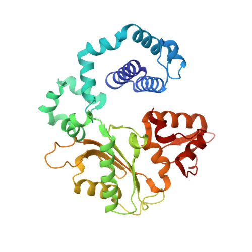
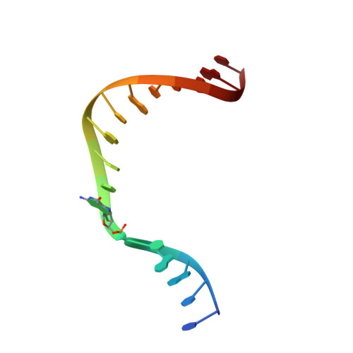
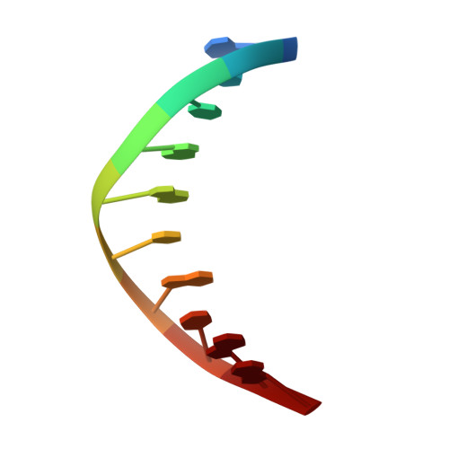
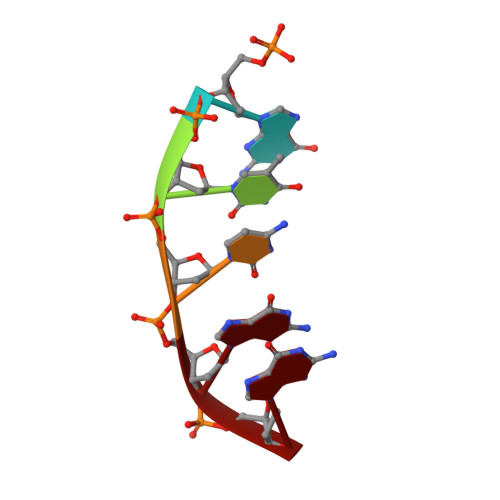
Metal-dependent conformational activation explains highly promutagenic replication across O6-methylguanine by human DNA polymerase beta.

Koag, M.C.Lee, S.

(2014) J Am Chem Soc 136: 5709-5721

  • DOI: https://doi.org/10.1021/ja500172d
  • Primary Citation of Related Structures:  
    4MF2, 4MFC, 4MFF, 4NXZ, 4NY8

  • PubMed Abstract: 

    Human DNA polymerase β (polβ) inserts, albeit slowly, T opposite the carcinogenic lesion O6-methylguanine (O6MeG) ∼30-fold more frequently than C. To gain insight into this promutagenic process, we solved four ternary structures of polβ with an incoming dCTP or dTTP analogue base-paired with O6MeG in the presence of active-site Mg(2+) or Mn(2+). The Mg(2+)-bound structures show that both the O6MeG·dCTP/dTTP-Mg(2+) complexes adopt an open protein conformation, staggered base pair, and one active-site metal ion. The Mn(2+)-bound structures reveal that, whereas the O6Me·dCTP-Mn(2+) complex assumes the similar altered conformation, the O6MeG·dTTP-Mn(2+) complex adopts a catalytically competent state with a closed protein conformation and pseudo-Watson-Crick base pair. On the basis of these observations, we conclude that polβ slows nucleotide incorporation opposite O6MeG by inducing an altered conformation suboptimal for catalysis and promotes mutagenic replication by allowing Watson-Crick-mode for O6MeG·T but not for O6MeG·C in the enzyme active site. The O6MeG·dTTP-Mn(2+) ternary structure, which represents the first structure of mismatched polβ ternary complex with a closed protein conformation and coplanar base pair, the first structure of pseudo-Watson-Crick O6MeG·T formed in the active site of a DNA polymerase, and a rare, if not the first, example of metal-dependent conformational activation of a DNA polymerase, indicate that catalytic metal-ion coordination is utilized as a kinetic checkpoint by polβ and is crucial for the conformational activation of polβ. Overall, our structural studies not only explain the promutagenic polβ catalysis across O6MeG but also provide new insights into the replication fidelity of polβ.


  • Organizational Affiliation

    Division of Medicinal Chemistry, College of Pharmacy, The University of Texas at Austin , Austin, Texas 78712, United States.


Macromolecules

Find similar proteins by:  (by identity cutoff)  |  3D Structure
Entity ID: 4
MoleculeChains Sequence LengthOrganismDetailsImage
DNA polymerase betaD [auth A]326Homo sapiensMutation(s): 0 
Gene Names: POLB
EC: 2.7.7.7 (PDB Primary Data), 4.2.99 (PDB Primary Data)
UniProt & NIH Common Fund Data Resources
Find proteins for P06746 (Homo sapiens)
Explore P06746 
Go to UniProtKB:  P06746
PHAROS:  P06746
GTEx:  ENSG00000070501 
Entity Groups  
Sequence Clusters30% Identity50% Identity70% Identity90% Identity95% Identity100% Identity
UniProt GroupP06746
Sequence Annotations
Expand
  • Reference Sequence

Find similar nucleic acids by:  Sequence   |   3D Structure  

Entity ID: 1
MoleculeChains LengthOrganismImage
5'-D(*CP*CP*GP*AP*CP*(6OG)P*TP*CP*GP*CP*AP*TP*CP*AP*GP*C)-3'A [auth T]16N/A
Sequence Annotations
Expand
  • Reference Sequence

Find similar nucleic acids by:  Sequence   |   3D Structure  

Entity ID: 2
MoleculeChains LengthOrganismImage
5'-D(*GP*CP*TP*GP*AP*TP*GP*CP*GP*A)-3'B [auth P]10N/A
Sequence Annotations
Expand
  • Reference Sequence

Find similar nucleic acids by:  Sequence   |   3D Structure  

Entity ID: 3
MoleculeChains LengthOrganismImage
5'-D(P*GP*TP*CP*GP*G)-3'C [auth D]5N/A
Sequence Annotations
Expand
  • Reference Sequence
Experimental Data & Validation

Experimental Data

  • Method: X-RAY DIFFRACTION
  • Resolution: 2.56 Å
  • R-Value Free: 0.255 
  • R-Value Work: 0.190 
  • R-Value Observed: 0.193 
  • Space Group: P 1 21 1
Unit Cell:
Length ( Å )Angle ( ˚ )
a = 50.803α = 90
b = 79.842β = 107.05
c = 55.442γ = 90
Software Package:
Software NamePurpose
PHENIXrefinement
PDB_EXTRACTdata extraction
StructureStudiodata collection
DENZOdata reduction
HKL-2000data scaling
MOLREPphasing
REFMACrefinement

Structure Validation

View Full Validation Report


Ligand Structure Quality Assessment 


Entry History 

Deposition Data

Revision History  (Full details and data files)

  • Version 1.0: 2014-04-16
    Type: Initial release
  • Version 1.1: 2014-07-02
    Changes: Database references
  • Version 1.2: 2019-07-17
    Changes: Data collection, Derived calculations, Refinement description
  • Version 1.3: 2024-02-28
    Changes: Data collection, Database references, Derived calculations