4NN0

Crystal structure of the C1QTNF5 globular domain in space group P63


Experimental Data Snapshot

  • Method: X-RAY DIFFRACTION
  • Resolution: 1.42 Å
  • R-Value Free: 0.142 
  • R-Value Work: 0.104 
  • R-Value Observed: 0.106 

wwPDB Validation   3D Report Full Report


This is version 1.2 of the entry. See complete history


Literature

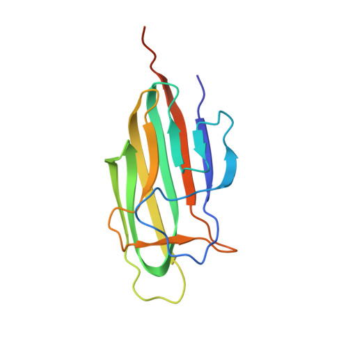
The macular degeneration-linked C1QTNF5 (S163) mutation causes higher-order structural rearrangements.

Tu, X.Palczewski, K.

(2014) J Struct Biol 186: 86-94

  • DOI: https://doi.org/10.1016/j.jsb.2014.02.001
  • Primary Citation of Related Structures:  
    4NN0

  • PubMed Abstract: 

    The C1q-tumor necrosis factor 5 (C1QTNF5) protein plays a significant role in retinal pigmented epithelium (RPE) cellular adhesion. The C1QTNF5 gene is co-transcribed with the frizzled-related protein (MFRP) gene. A Ser-to-Arg mutation at site 163 (S163R) in C1QTNF5 is known to cause late-onset retinal macular degeneration (L-ORMD). Here we also found that C1QTNF5 monomers can multimerize into a bouquet-like octadecamer. We found that a novel intermolecular hydrogen-bond network of S163 that glues adjacent globular heads of C1QTNF5 together was weakened or abolished by the R163 pathogenic mutation. These findings could underlie the structural basis of this protein's adhesive function and relate to the pathogenesis of its S163R mutation. Additionally, the fact that C1QTNF5 immobilized to a resin selectively enriched detergent extracted membrane-bound MFRP, further confirmed their interaction, implying functions other than cellular adhesion for C1QTNF5.


  • Organizational Affiliation

    Department of Pharmacology, School of Medicine, Case Western Reserve University, USA.


Macromolecules
Find similar proteins by:  (by identity cutoff)  |  3D Structure
Entity ID: 1
MoleculeChains Sequence LengthOrganismDetailsImage
Complement C1q tumor necrosis factor-related protein 5
A, B, C
148Homo sapiensMutation(s): 0 
Gene Names: C1QTNF5CTRP5UNQ303/PRO344
UniProt & NIH Common Fund Data Resources
Find proteins for Q9BXJ0 (Homo sapiens)
Explore Q9BXJ0 
Go to UniProtKB:  Q9BXJ0
PHAROS:  Q9BXJ0
GTEx:  ENSG00000223953 
Entity Groups  
Sequence Clusters30% Identity50% Identity70% Identity90% Identity95% Identity100% Identity
UniProt GroupQ9BXJ0
Sequence Annotations
Expand
  • Reference Sequence
Small Molecules
Ligands 3 Unique
IDChains Name / Formula / InChI Key2D Diagram3D Interactions
SO4
Query on SO4

Download Ideal Coordinates CCD File 
P [auth A],
W [auth B],
X [auth B]
SULFATE ION
O4 S
QAOWNCQODCNURD-UHFFFAOYSA-L
EDO
Query on EDO

Download Ideal Coordinates CCD File 
D [auth A]
E [auth A]
F [auth A]
G [auth A]
H [auth A]
D [auth A],
E [auth A],
F [auth A],
G [auth A],
H [auth A],
I [auth A],
J [auth A],
K [auth A],
L [auth A],
M [auth A],
N [auth A],
O [auth A],
R [auth B],
S [auth B],
T [auth B],
U [auth B],
V [auth B],
Z [auth C]
1,2-ETHANEDIOL
C2 H6 O2
LYCAIKOWRPUZTN-UHFFFAOYSA-N
ACT
Query on ACT

Download Ideal Coordinates CCD File 
Q [auth A],
Y [auth B]
ACETATE ION
C2 H3 O2
QTBSBXVTEAMEQO-UHFFFAOYSA-M
Experimental Data & Validation

Experimental Data

  • Method: X-RAY DIFFRACTION
  • Resolution: 1.42 Å
  • R-Value Free: 0.142 
  • R-Value Work: 0.104 
  • R-Value Observed: 0.106 
  • Space Group: P 63
Unit Cell:
Length ( Å )Angle ( ˚ )
a = 50.603α = 90
b = 50.603β = 90
c = 268.531γ = 120
Software Package:
Software NamePurpose
ADSCdata collection
PHASERphasing
REFMACrefinement
DPSdata reduction

Structure Validation

View Full Validation Report


Entry History 

Deposition Data

Revision History  (Full details and data files)

  • Version 1.0: 2014-03-26
    Type: Initial release
  • Version 1.1: 2014-04-23
    Changes: Database references
  • Version 1.2: 2023-09-20
    Changes: Data collection, Database references, Derived calculations, Refinement description