4NC9

Crystal structure of phosphatidyl mannosyltransferase PimA


Experimental Data Snapshot

  • Method: X-RAY DIFFRACTION
  • Resolution: 3.19 Å
  • R-Value Free: 0.256 
  • R-Value Work: 0.212 
  • R-Value Observed: 0.214 

wwPDB Validation   3D Report Full Report


This is version 1.2 of the entry. See complete history


Literature

Secondary structure reshuffling modulates glycosyltransferase function at the membrane.

Giganti, D.Albesa-Jove, D.Urresti, S.Rodrigo-Unzueta, A.Martinez, M.A.Comino, N.Barilone, N.Bellinzoni, M.Chenal, A.Guerin, M.E.Alzari, P.M.

(2015) Nat Chem Biol 11: 16-18

  • DOI: https://doi.org/10.1038/nchembio.1694
  • Primary Citation of Related Structures:  
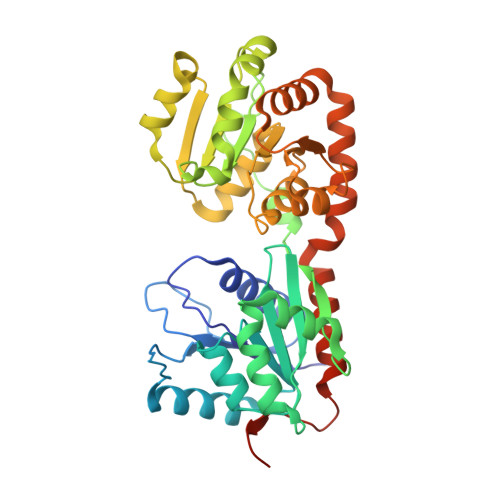
    4N9W, 4NC9

  • PubMed Abstract: 

    Secondary structure refolding is a key event in biology as it modulates the conformation of many proteins in the cell, generating functional or aberrant states. The crystal structures of mannosyltransferase PimA reveal an exceptional flexibility of the protein along the catalytic cycle, including β-strand-to-α-helix and α-helix-to-β-strand transitions. These structural changes modulate catalysis and are promoted by interactions of the protein with anionic phospholipids in the membrane.


  • Organizational Affiliation

    1] Institut Pasteur, Unité de Microbiologie Structurale, CNRS UMR 3528, Paris, France. [2] Unidad de Biofisica, Centro Mixto Consejo Superior de Investigaciones Cientificas-Universidad del País Vasco/Euskal Herriko Unibertsitatea (CSIC,UPV/EHU), Leioa, Bizkaia, Spain. [3] Departamento de Bioquímica, Universidad del País Vasco, Spain.


Macromolecules
Find similar proteins by:  (by identity cutoff)  |  3D Structure
Entity ID: 1
MoleculeChains Sequence LengthOrganismDetailsImage
GDP-mannose-dependent alpha-(1-2)-phosphatidylinositol mannosyltransferase
A, B, C, D
390Mycolicibacterium smegmatis MC2 155Mutation(s): 0 
Gene Names: MSMEG_2935MSMEI_2861pimA
EC: 2.4.1.57
Membrane Entity: Yes 
UniProt
Find proteins for A0QWG6 (Mycolicibacterium smegmatis (strain ATCC 700084 / mc(2)155))
Explore A0QWG6 
Go to UniProtKB:  A0QWG6
Entity Groups  
Sequence Clusters30% Identity50% Identity70% Identity90% Identity95% Identity100% Identity
UniProt GroupA0QWG6
Sequence Annotations
Expand
  • Reference Sequence
Experimental Data & Validation

Experimental Data

  • Method: X-RAY DIFFRACTION
  • Resolution: 3.19 Å
  • R-Value Free: 0.256 
  • R-Value Work: 0.212 
  • R-Value Observed: 0.214 
  • Space Group: P 32 2 1
Unit Cell:
Length ( Å )Angle ( ˚ )
a = 137.62α = 90
b = 137.62β = 90
c = 168.641γ = 120
Software Package:
Software NamePurpose
ADSCdata collection
PHASERphasing
PHENIXrefinement
XDSdata reduction
SCALAdata scaling

Structure Validation

View Full Validation Report


Entry History 

Deposition Data

Revision History  (Full details and data files)

  • Version 1.0: 2014-11-12
    Type: Initial release
  • Version 1.1: 2015-06-10
    Changes: Database references
  • Version 1.2: 2023-09-20
    Changes: Data collection, Database references, Refinement description