4MRQ

Crystal Structure of wild-type unphosphorylated PMM/PGM


Experimental Data Snapshot

  • Method: X-RAY DIFFRACTION
  • Resolution: 1.90 Å
  • R-Value Free: 0.205 
  • R-Value Work: 0.165 
  • R-Value Observed: 0.167 

wwPDB Validation   3D Report Full Report


This is version 1.2 of the entry. See complete history


Literature

Promotion of enzyme flexibility by dephosphorylation and coupling to the catalytic mechanism of a phosphohexomutase.

Lee, Y.Villar, M.T.Artigues, A.Beamer, L.J.

(2014) J Biol Chem 289: 4674-4682

  • DOI: https://doi.org/10.1074/jbc.M113.532226
  • Primary Citation of Related Structures:  
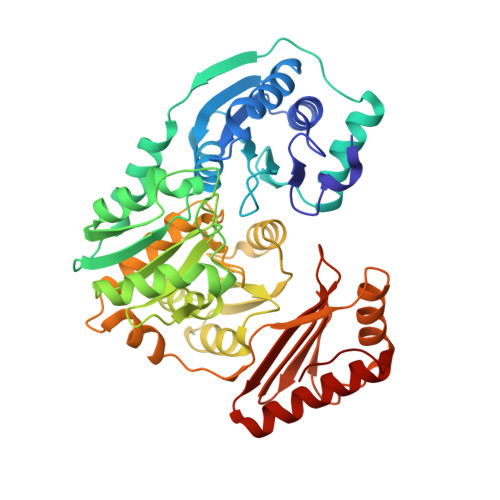
    4MRQ

  • PubMed Abstract: 

    The enzyme phosphomannomutase/phosphoglucomutase (PMM/PGM) from Pseudomonas aeruginosa catalyzes an intramolecular phosphoryl transfer across its phosphosugar substrates, which are precursors in the synthesis of exoproducts involved in bacterial virulence. Previous structural studies of PMM/PGM have established a key role for conformational change in its multistep reaction, which requires a dramatic 180° reorientation of the intermediate within the active site. Here hydrogen-deuterium exchange by mass spectrometry and small angle x-ray scattering were used to probe the conformational flexibility of different forms of PMM/PGM in solution, including its active, phosphorylated state and the unphosphorylated state that occurs transiently during the catalytic cycle. In addition, the effects of ligand binding were assessed through use of a substrate analog. We found that both phosphorylation and binding of ligand produce significant effects on deuterium incorporation. Phosphorylation of the conserved catalytic serine has broad effects on residues in multiple domains and is supported by small angle x-ray scattering data showing that the unphosphorylated enzyme is less compact in solution. The effects of ligand binding are generally manifested near the active site cleft and at a domain interface that is a site of conformational change. These results suggest that dephosphorylation of the enzyme may play two critical functional roles: a direct role in the chemical step of phosphoryl transfer and secondly through propagation of structural flexibility. We propose a model whereby increased enzyme flexibility facilitates the reorientation of the reaction intermediate, coupling changes in structural dynamics with the unique catalytic mechanism of this enzyme.


  • Organizational Affiliation

    From the Chemistry and.


Macromolecules
Find similar proteins by:  (by identity cutoff)  |  3D Structure
Entity ID: 1
MoleculeChains Sequence LengthOrganismDetailsImage
Phosphomannomutase/phosphoglucomutase455Pseudomonas aeruginosa PAO1Mutation(s): 0 
Gene Names: algCPA5322
EC: 5.4.2.2 (PDB Primary Data), 5.4.2.8 (PDB Primary Data)
UniProt
Find proteins for P26276 (Pseudomonas aeruginosa (strain ATCC 15692 / DSM 22644 / CIP 104116 / JCM 14847 / LMG 12228 / 1C / PRS 101 / PAO1))
Explore P26276 
Go to UniProtKB:  P26276
Entity Groups  
Sequence Clusters30% Identity50% Identity70% Identity90% Identity95% Identity100% Identity
UniProt GroupP26276
Sequence Annotations
Expand
  • Reference Sequence
Small Molecules
Ligands 5 Unique
IDChains Name / Formula / InChI Key2D Diagram3D Interactions
PGE
Query on PGE

Download Ideal Coordinates CCD File 
J [auth A],
K [auth A],
L [auth A]
TRIETHYLENE GLYCOL
C6 H14 O4
ZIBGPFATKBEMQZ-UHFFFAOYSA-N
TLA
Query on TLA

Download Ideal Coordinates CCD File 
C [auth A]L(+)-TARTARIC ACID
C4 H6 O6
FEWJPZIEWOKRBE-JCYAYHJZSA-N
PEG
Query on PEG

Download Ideal Coordinates CCD File 
E [auth A],
F [auth A],
G [auth A],
H [auth A],
I [auth A]
DI(HYDROXYETHYL)ETHER
C4 H10 O3
MTHSVFCYNBDYFN-UHFFFAOYSA-N
ZN
Query on ZN

Download Ideal Coordinates CCD File 
B [auth A]ZINC ION
Zn
PTFCDOFLOPIGGS-UHFFFAOYSA-N
EDO
Query on EDO

Download Ideal Coordinates CCD File 
D [auth A],
M [auth A],
N [auth A],
O [auth A]
1,2-ETHANEDIOL
C2 H6 O2
LYCAIKOWRPUZTN-UHFFFAOYSA-N
Experimental Data & Validation

Experimental Data

  • Method: X-RAY DIFFRACTION
  • Resolution: 1.90 Å
  • R-Value Free: 0.205 
  • R-Value Work: 0.165 
  • R-Value Observed: 0.167 
  • Space Group: P 21 21 21
Unit Cell:
Length ( Å )Angle ( ˚ )
a = 70.541α = 90
b = 72.003β = 90
c = 92.349γ = 90
Software Package:
Software NamePurpose
REFMACrefinement
PDB_EXTRACTdata extraction

Structure Validation

View Full Validation Report


Entry History 

Deposition Data

Revision History  (Full details and data files)

  • Version 1.0: 2014-01-08
    Type: Initial release
  • Version 1.1: 2014-11-05
    Changes: Database references
  • Version 1.2: 2023-09-20
    Changes: Data collection, Database references, Derived calculations, Refinement description