4MFE

Structure of the carboxyl transferase domain from Rhizobium etli pyruvate carboxylase with 3-hydroxypyruvate


Experimental Data Snapshot

  • Method: X-RAY DIFFRACTION
  • Resolution: 2.61 Å
  • R-Value Free: 0.225 
  • R-Value Work: 0.177 
  • R-Value Observed: 0.179 

wwPDB Validation   3D Report Full Report


Ligand Structure Quality Assessment 


This is version 1.6 of the entry. See complete history


Literature

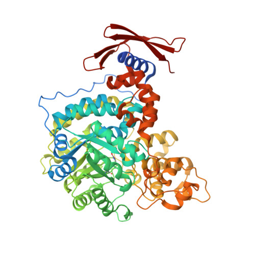
Insights into the carboxyltransferase reaction of pyruvate carboxylase from the structures of bound product and intermediate analogs.

Lietzan, A.D.St.Maurice, M.

(2013) Biochem Biophys Res Commun 441: 377-382

  • DOI: https://doi.org/10.1016/j.bbrc.2013.10.066
  • Primary Citation of Related Structures:  
    4MFD, 4MFE, 4MIM

  • PubMed Abstract: 

    Pyruvate carboxylase (PC) is a biotin-dependent enzyme that catalyzes the MgATP- and bicarbonate-dependent carboxylation of pyruvate to oxaloacetate, an important anaplerotic reaction in central metabolism. The carboxyltransferase (CT) domain of PC catalyzes the transfer of a carboxyl group from carboxybiotin to the accepting substrate, pyruvate. It has been hypothesized that the reactive enolpyruvate intermediate is stabilized through a bidentate interaction with the metal ion in the CT domain active site. Whereas bidentate ligands are commonly observed in enzymes catalyzing reactions proceeding through an enolpyruvate intermediate, no bidentate interaction has yet been observed in the CT domain of PC. Here, we report three X-ray crystal structures of the Rhizobium etli PC CT domain with the bound inhibitors oxalate, 3-hydroxypyruvate, and 3-bromopyruvate. Oxalate, a stereoelectronic mimic of the enolpyruvate intermediate, does not interact directly with the metal ion. Instead, oxalate is buried in a pocket formed by several positively charged amino acid residues and the metal ion. Furthermore, both 3-hydroxypyruvate and 3-bromopyruvate, analogs of the reaction product oxaloacetate, bind in an identical manner to oxalate suggesting that the substrate maintains its orientation in the active site throughout catalysis. Together, these structures indicate that the substrates, products and intermediates in the PC-catalyzed reaction are not oriented in the active site as previously assumed. The absence of a bidentate interaction with the active site metal appears to be a unique mechanistic feature among the small group of biotin-dependent enzymes that act on α-keto acid substrates.


  • Organizational Affiliation

    Department of Biological Sciences, Marquette University, Milwaukee, WI 53201, USA.


Macromolecules
Find similar proteins by:  (by identity cutoff)  |  3D Structure
Entity ID: 1
MoleculeChains Sequence LengthOrganismDetailsImage
PYRUVATE CARBOXYLASE
A, B, C, D
632Rhizobium etli CFN 42Mutation(s): 0 
Gene Names: pycRHE_CH04002
EC: 6.4.1.1
UniProt
Find proteins for Q2K340 (Rhizobium etli (strain CFN 42 / ATCC 51251))
Explore Q2K340 
Go to UniProtKB:  Q2K340
Entity Groups  
Sequence Clusters30% Identity50% Identity70% Identity90% Identity95% Identity100% Identity
UniProt GroupQ2K340
Sequence Annotations
Expand
  • Reference Sequence
Small Molecules
Ligands 6 Unique
IDChains Name / Formula / InChI Key2D Diagram3D Interactions
BTN
Query on BTN

Download Ideal Coordinates CCD File 
G [auth A],
M [auth B],
R [auth C],
X [auth D]
BIOTIN
C10 H16 N2 O3 S
YBJHBAHKTGYVGT-ZKWXMUAHSA-N
3PY
Query on 3PY

Download Ideal Coordinates CCD File 
F [auth A],
L [auth B],
Q [auth C],
W [auth D]
3-HYDROXYPYRUVIC ACID
C3 H4 O4
HHDDCCUIIUWNGJ-UHFFFAOYSA-N
GOL
Query on GOL

Download Ideal Coordinates CCD File 
J [auth A],
U [auth D]
GLYCEROL
C3 H8 O3
PEDCQBHIVMGVHV-UHFFFAOYSA-N
ZN
Query on ZN

Download Ideal Coordinates CCD File 
E [auth A],
K [auth B],
P [auth C],
V [auth D]
ZINC ION
Zn
PTFCDOFLOPIGGS-UHFFFAOYSA-N
CL
Query on CL

Download Ideal Coordinates CCD File 
I [auth A],
O [auth B],
T [auth C]
CHLORIDE ION
Cl
VEXZGXHMUGYJMC-UHFFFAOYSA-M
MG
Query on MG

Download Ideal Coordinates CCD File 
H [auth A],
N [auth B],
S [auth C],
Y [auth D]
MAGNESIUM ION
Mg
JLVVSXFLKOJNIY-UHFFFAOYSA-N
Modified Residues  1 Unique
IDChains TypeFormula2D DiagramParent
KCX
Query on KCX
A, B, C, D
L-PEPTIDE LINKINGC7 H14 N2 O4LYS
Binding Affinity Annotations 
IDSourceBinding Affinity
3PY PDBBind:  4MFE Ki: 5.40e+6 (nM) from 1 assay(s)
Experimental Data & Validation

Experimental Data

  • Method: X-RAY DIFFRACTION
  • Resolution: 2.61 Å
  • R-Value Free: 0.225 
  • R-Value Work: 0.177 
  • R-Value Observed: 0.179 
  • Space Group: P 21 21 21
Unit Cell:
Length ( Å )Angle ( ˚ )
a = 84.254α = 90
b = 157.849β = 90
c = 243.319γ = 90
Software Package:
Software NamePurpose
MD2data collection
PHASERphasing
REFMACrefinement
DENZOdata reduction
SCALEPACKdata scaling

Structure Validation

View Full Validation Report


Ligand Structure Quality Assessment 


Entry History 

Deposition Data

Revision History  (Full details and data files)

  • Version 1.0: 2013-11-13
    Type: Initial release
  • Version 1.1: 2013-12-04
    Changes: Database references
  • Version 1.2: 2017-11-22
    Changes: Database references
  • Version 1.3: 2018-01-24
    Changes: Database references
  • Version 1.4: 2018-01-31
    Changes: Database references, Structure summary
  • Version 1.5: 2023-09-20
    Changes: Data collection, Database references, Derived calculations, Refinement description
  • Version 1.6: 2023-12-06
    Changes: Data collection