4M7H

Crystal structure of tetrameric fibrinogen-like recognition domain of FIBCD1


Experimental Data Snapshot

  • Method: X-RAY DIFFRACTION
  • Resolution: 2.00 Å
  • R-Value Free: 0.209 
  • R-Value Work: 0.183 
  • R-Value Observed: 0.192 

wwPDB Validation   3D Report Full Report


Ligand Structure Quality Assessment 


This is version 1.4 of the entry. See complete history


Literature

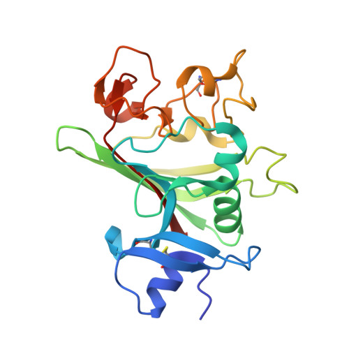
Crystal Structure of the Tetrameric Fibrinogen-like Recognition Domain of Fibrinogen C Domain Containing 1 (FIBCD1) Protein.

Shrive, A.K.Moeller, J.B.Burns, I.Paterson, J.M.Shaw, A.J.Schlosser, A.Sorensen, G.L.Greenhough, T.J.Holmskov, U.

(2014) J Biol Chem 289: 2880-2887

  • DOI: https://doi.org/10.1074/jbc.M113.520577
  • Primary Citation of Related Structures:  
    4M7F, 4M7H

  • PubMed Abstract: 

    The high resolution crystal structures of a recombinant fragment of the C-terminal fibrinogen-like recognition domain of FIBCD1, a vertebrate receptor that binds chitin, have been determined. The overall tetrameric structure shows similarity in structure and aggregation to the horseshoe crab innate immune protein tachylectin 5A. The high affinity ligand N-acetylmannosamine (ManNAc) binds in the S1 site, predominantly via the acetyl group with the oxygen and acetamide nitrogen hydrogen-bonded to the protein and the methyl group inserted into a hydrophobic pocket. The binding of the ManNAc pyranose ring differs markedly between the two independent subunits, but in all structures the binding of the N-acetyl group is conserved. In the native structure, a crystal contact results in one of the independent protomers binding the first GlcNAc of the Asn(340) N-linked glycan on the other independent protomer. In the ligand-bound structure this GlcNAc is replaced by the higher affinity ligand ManNAc. In addition, a sulfate ion has been modeled into the electron density at a location similar to the S3 binding site in L-ficolin, whereas in the native structure an acetate ion has been placed in the S1 N-acetyl binding site, and a sulfate ion has been placed adjacent to this site. These ion binding sites are ideally placed to receive the N-acetyl and sulfate groups of sulfated GalNAc residues of glycosaminoglycans such as chondroitin and dermatan sulfate. Together, these structures give insight into important determinants of ligand selectivity, demonstrating versatility in recognition and binding while maintaining conservation in N-acetyl and calcium binding.


  • Organizational Affiliation

    From the Research Institute of Science and Technology in Medicine, School of Life Sciences, Keele University, Staffordshire ST5 5BG, United Kingdom and.


Macromolecules
Find similar proteins by:  (by identity cutoff)  |  3D Structure
Entity ID: 1
MoleculeChains Sequence LengthOrganismDetailsImage
Fibrinogen C domain-containing protein 1
A, B
226Homo sapiensMutation(s): 0 
Gene Names: FIBCD1UNQ701/PRO1346
UniProt & NIH Common Fund Data Resources
Find proteins for Q8N539 (Homo sapiens)
Explore Q8N539 
Go to UniProtKB:  Q8N539
PHAROS:  Q8N539
GTEx:  ENSG00000130720 
Entity Groups  
Sequence Clusters30% Identity50% Identity70% Identity90% Identity95% Identity100% Identity
UniProt GroupQ8N539
Sequence Annotations
Expand
  • Reference Sequence
Small Molecules
Ligands 5 Unique
IDChains Name / Formula / InChI Key2D Diagram3D Interactions
NAG
Query on NAG

Download Ideal Coordinates CCD File 
D [auth A]2-acetamido-2-deoxy-beta-D-glucopyranose
C8 H15 N O6
OVRNDRQMDRJTHS-FMDGEEDCSA-N
SO4
Query on SO4

Download Ideal Coordinates CCD File 
F [auth A],
G [auth A],
J [auth B]
SULFATE ION
O4 S
QAOWNCQODCNURD-UHFFFAOYSA-L
GOL
Query on GOL

Download Ideal Coordinates CCD File 
I [auth B]GLYCEROL
C3 H8 O3
PEDCQBHIVMGVHV-UHFFFAOYSA-N
ACY
Query on ACY

Download Ideal Coordinates CCD File 
E [auth A]ACETIC ACID
C2 H4 O2
QTBSBXVTEAMEQO-UHFFFAOYSA-N
CA
Query on CA

Download Ideal Coordinates CCD File 
C [auth A],
H [auth B]
CALCIUM ION
Ca
BHPQYMZQTOCNFJ-UHFFFAOYSA-N
Experimental Data & Validation

Experimental Data

  • Method: X-RAY DIFFRACTION
  • Resolution: 2.00 Å
  • R-Value Free: 0.209 
  • R-Value Work: 0.183 
  • R-Value Observed: 0.192 
  • Space Group: P 4
Unit Cell:
Length ( Å )Angle ( ˚ )
a = 118.56α = 90
b = 118.56β = 90
c = 44.25γ = 90
Software Package:
Software NamePurpose
MOSFLMdata reduction
SCALAdata scaling
AMoREphasing
CNSrefinement
PDB_EXTRACTdata extraction
ADSCdata collection

Structure Validation

View Full Validation Report


Ligand Structure Quality Assessment 


Entry History 

Deposition Data

Revision History  (Full details and data files)

  • Version 1.0: 2013-12-11
    Type: Initial release
  • Version 1.1: 2013-12-25
    Changes: Database references
  • Version 1.2: 2014-02-19
    Changes: Database references
  • Version 1.3: 2020-07-29
    Type: Remediation
    Reason: Carbohydrate remediation
    Changes: Data collection, Derived calculations, Structure summary
  • Version 1.4: 2023-09-20
    Changes: Data collection, Database references, Refinement description, Structure summary