4LMB

Crystal structure analysis of O-acetylserine sulfhydrylase CysK2 complexed with cystine from Microcystis aeruginosa 7806


Experimental Data Snapshot

  • Method: X-RAY DIFFRACTION
  • Resolution: 1.91 Å
  • R-Value Free: 0.230 
  • R-Value Work: 0.202 
  • R-Value Observed: 0.203 

wwPDB Validation   3D Report Full Report


Ligand Structure Quality Assessment 


This is version 1.2 of the entry. See complete history


Literature

Structural and biochemical analyses of Microcystis aeruginosa O-acetylserine sulfhydrylases reveal a negative feedback regulation of cysteine biosynthesis.

Lu, M.Xu, B.Y.Zhou, K.Cheng, W.Jiang, Y.L.Chen, Y.Zhou, C.Z.

(2014) Biochim Biophys Acta 1844: 308-315

  • DOI: https://doi.org/10.1016/j.bbapap.2013.11.008
  • Primary Citation of Related Structures:  
    4LMA, 4LMB

  • PubMed Abstract: 

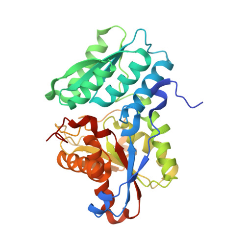
    O-acetylserine sulfhydrylase (OASS) catalyzes the final step of cysteine biosynthesis from O-acetylserine (OAS) and inorganic sulfide in plants and bacteria. Bioinformatics analyses combined with activity assays enabled us to annotate the two putative genes of Microcystis aeruginosa PCC 7806 to CysK1 and CysK2, which encode the two 75% sequence-identical OASS paralogs. Moreover, we solved the crystal structures of CysK1 at 2.30Ǻ and cystine-complexed CysK2 at 1.91Ǻ, revealing a quite similar overall structure that belongs to the family of fold-type II PLP-dependent enzymes. Structural comparison indicated a significant induced fit upon binding to the cystine, which occupies the binding site for the substrate OAS and blocks the product release tunnel. Subsequent enzymatic assays further confirmed that cystine is a competitive inhibitor of the substrate OAS. Moreover, multiple-sequence alignment revealed that the cystine-binding residues are highly conserved in all OASS proteins, suggesting that this auto-inhibition of cystine might be a universal mechanism of cysteine biosynthesis pathway.


  • Organizational Affiliation

    Hefei National Laboratory for Physical Sciences at the Microscale and School of Life Sciences, University of Science and Technology of China, Hefei, Anhui 230027, People's Republic of China.


Macromolecules
Find similar proteins by:  (by identity cutoff)  |  3D Structure
Entity ID: 1
MoleculeChains Sequence LengthOrganismDetailsImage
Cysteine synthase322Microcystis aeruginosa PCC 7806Mutation(s): 0 
Gene Names: cysKIPF_2058IPF_3084
EC: 2.5.1.47
UniProt
Find proteins for A8YBP8 (Microcystis aeruginosa (strain PCC 7806))
Explore A8YBP8 
Go to UniProtKB:  A8YBP8
Entity Groups  
Sequence Clusters30% Identity50% Identity70% Identity90% Identity95% Identity100% Identity
UniProt GroupA8YBP8
Sequence Annotations
Expand
  • Reference Sequence
Experimental Data & Validation

Experimental Data

  • Method: X-RAY DIFFRACTION
  • Resolution: 1.91 Å
  • R-Value Free: 0.230 
  • R-Value Work: 0.202 
  • R-Value Observed: 0.203 
  • Space Group: P 65 2 2
Unit Cell:
Length ( Å )Angle ( ˚ )
a = 99.846α = 90
b = 99.846β = 90
c = 164.141γ = 120
Software Package:
Software NamePurpose
ADSCdata collection
AMoREphasing
REFMACrefinement
HKL-2000data reduction
SCALAdata scaling

Structure Validation

View Full Validation Report


Ligand Structure Quality Assessment 


Entry History 

Deposition Data

  • Released Date: 2014-06-04 
  • Deposition Author(s): Lu, M.

Revision History  (Full details and data files)

  • Version 1.0: 2014-06-04
    Type: Initial release
  • Version 1.1: 2023-11-08
    Changes: Data collection, Database references, Derived calculations, Refinement description
  • Version 1.2: 2023-11-15
    Changes: Derived calculations