Structural basis of the chiral selectivity of Pseudomonas cepacia lipase
Lang, D.A., Mannesse, M.L.M., De Haas, G., Verheij, H.M., Dijkstra, B.W.(1998) Eur J Biochem 254: 333-340
- PubMed: 9660188
- DOI: https://doi.org/10.1046/j.1432-1327.1998.2540333.x
- Primary Citation of Related Structures:
4LIP, 5LIP - PubMed Abstract:
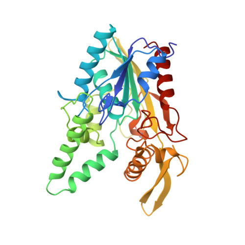
To investigate the enantioselectivity of Pseudomonas cepacia lipase, inhibition studies were performed with Sc- and Rc-(Rp,Sp)-1,2-dialkylcarbamoylglycero-3-O-p-nitrophenyl alkylphosphonates of different alkyl chain lengths. P. cepacia lipase was most rapidly inactivated by Rc-(Rp,Sp)-1,2-dioctylcarbamoylglycero-3-O-p-nitrophenyl octylphosphonate (Rc-trioctyl) with an inactivation half-time of 75 min, while that for the Sc-(Rp,Sp)-1,2-dioctylcarbamoylglycero-3-O-p-nitrophenyl octyl-phosphonate (Sc-trioctyl) compound was 530 min. X-ray structures were obtained of P. cepacia lipase after reaction with Rc-trioctyl to 0.29-nm resolution at pH 4 and covalently modified with Rc-(Rp,Sp)-1,2-dibutylcarbamoylglycero-3-O-p-nitrophenyl butyl-phosphonate (Rc-tributyl) to 0.175-nm resolution at pH 8.5. The three-dimensional structures reveal that both triacylglycerol analogues had reacted with the active-site Ser87, forming a covalent complex. The bound phosphorus atom shows the same chirality (Sp) in both complexes despite the use of a racemic (Rp,Sp) mixture at the phosphorus atom of the triacylglycerol analogues. In the structure of Rc-tributyl-complexed P. cepacia lipase, the diacylglycerol moiety has been lost due to an aging reaction, and only the butyl phosphonate remains visible in the electron density. In the Rc-trioctyl complex the complete inhibitor is clearly defined; it adopts a bent tuning fork conformation. Unambiguously, four binding pockets for the triacylglycerol could be detected: an oxyanion hole and three pockets which accommodate the sn-1, sn-2, and sn-3 fatty acid chains. Van der Waals' interactions are the main forces that keep the radyl groups of the triacylglycerol analogue in position and, in addition, a hydrogen bond to the carbonyl oxygen of the sn-2 chain contributes to fixing the position of the inhibitor.
Organizational Affiliation:
BIOSON Research Institute and Laboratory of Biophysical Chemistry, University of Groningen, The Netherlands.