4L62

Crystal Structure of Pseudomonas aeruginosa transcriptional regulator PA2196 bound to its operator DNA


Experimental Data Snapshot

  • Method: X-RAY DIFFRACTION
  • Resolution: 2.90 Å
  • R-Value Free: 0.238 
  • R-Value Work: 0.198 
  • R-Value Observed: 0.200 

wwPDB Validation   3D Report Full Report


This is version 1.2 of the entry. See complete history


Literature

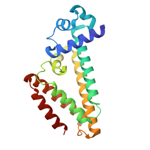
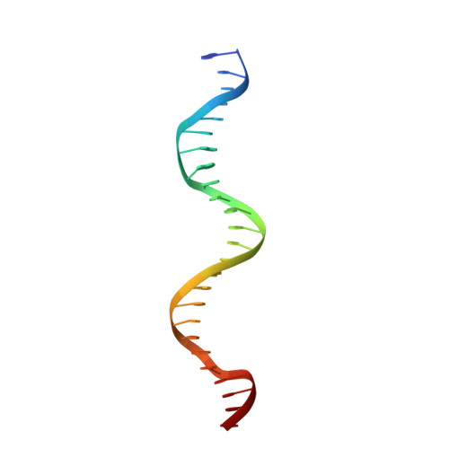
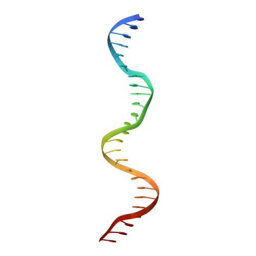
Crystal structure of Pseudomonas aeruginosa transcriptional regulator PA2196 bound to its operator DNA.

Kim, Y.Kang, Y.Choe, J.

(2013) Biochem Biophys Res Commun 440: 317-321

  • DOI: https://doi.org/10.1016/j.bbrc.2013.09.074
  • Primary Citation of Related Structures:  
    4L62

  • PubMed Abstract: 

    Pseudomonas aeruginosa is a major opportunistic human pathogen. PA2196 from P. aeruginosa is a member of TetR family of transcriptional repressors, which is involved in adaptation to environmental changes as well as bacterial antibiotic resistance. PA2196 consists of nine α-helical bundles divided into two separate domains. The N-terminal domain, called the DNA-binding domain, is composed of helices α1-α3 and has a helix-turn-helix motif. The C-terminal domain, called the ligand-binding domain, has a hydrophobic pocket for ligand binding. Here, PA2196 was shown to bind to a 25 bp semi-palindromic dsDNA located in the upstream region of its own gene. The crystal structure of the PA2196-25mer dsDNA complex determined at a resolution of 2.9 Å revealed that two dimers of PA2196 bound to one dsDNA, with each monomer interacting with the major groove of DNA. Especially, residues in helix α3, including Lys41, Gly42, Ser43, and Tyr45, interacted mainly with the base and phosphate backbone of dsDNA. PA2196 underwent large conformational changes upon DNA binding, as the distances between DNA-binding domains measured between two G42s in subunits A and B decreased from 41.7 Å to 36.8 Å. Our crystal structure of PA2196-25mer dsDNA complex revealed that PA2196 is similar to QacR in that two dimers bound to one dsDNA through specific interactions.


  • Organizational Affiliation

    Department of Life Science, University of Seoul, Seoul 130-743, Republic of Korea.


Macromolecules

Find similar proteins by:  (by identity cutoff)  |  3D Structure
Entity ID: 1
MoleculeChains Sequence LengthOrganismDetailsImage
Transcriptional regulator
A, B, C, D, E
A, B, C, D, E, F, G, H, I, J, K, L, M, N, O, P
190Pseudomonas aeruginosa PAO1Mutation(s): 0 
Gene Names: PA2196
UniProt
Find proteins for Q9I1S1 (Pseudomonas aeruginosa (strain ATCC 15692 / DSM 22644 / CIP 104116 / JCM 14847 / LMG 12228 / 1C / PRS 101 / PAO1))
Explore Q9I1S1 
Go to UniProtKB:  Q9I1S1
Entity Groups  
Sequence Clusters30% Identity50% Identity70% Identity90% Identity95% Identity100% Identity
UniProt GroupQ9I1S1
Sequence Annotations
Expand
  • Reference Sequence

Find similar nucleic acids by:  Sequence   |   3D Structure  

Entity ID: 2
MoleculeChains LengthOrganismImage
DNA (25-MER)
Q, S, U, W
25N/A
Sequence Annotations
Expand
  • Reference Sequence

Find similar nucleic acids by:  Sequence   |   3D Structure  

Entity ID: 3
MoleculeChains LengthOrganismImage
DNA (25-MER)
R, T, V, X
25N/A
Sequence Annotations
Expand
  • Reference Sequence
Experimental Data & Validation

Experimental Data

  • Method: X-RAY DIFFRACTION
  • Resolution: 2.90 Å
  • R-Value Free: 0.238 
  • R-Value Work: 0.198 
  • R-Value Observed: 0.200 
  • Space Group: P 32 2 1
Unit Cell:
Length ( Å )Angle ( ˚ )
a = 211.994α = 90
b = 211.994β = 90
c = 282.773γ = 120
Software Package:
Software NamePurpose
HKL-2000data collection
PHENIXmodel building
PHENIXrefinement
HKL-2000data reduction
HKL-2000data scaling
PHENIXphasing

Structure Validation

View Full Validation Report


Entry History 

Deposition Data

Revision History  (Full details and data files)

  • Version 1.0: 2013-10-23
    Type: Initial release
  • Version 1.1: 2013-11-06
    Changes: Database references
  • Version 1.2: 2024-02-28
    Changes: Data collection, Database references