4KRV

Crystal structure of catalytic domain of bovine beta1,4-galactosyltransferase mutant M344H-GalT1 complex with 6-sulfo-GlcNAc


Experimental Data Snapshot

  • Method: X-RAY DIFFRACTION
  • Resolution: 2.40 Å
  • R-Value Free: 0.249 
  • R-Value Work: 0.188 
  • R-Value Observed: 0.191 

wwPDB Validation   3D Report Full Report


Ligand Structure Quality Assessment 


This is version 2.1 of the entry. See complete history


Literature

Investigations on beta 1,4-galactosyltransferase I using 6-sulfo-GlcNAc as an acceptor sugar substrate.

Ramakrishnan, B.Moncrief, A.J.Davis, T.A.Holland, L.A.Qasba, P.K.

(2013) Glycoconj J 30: 835-842

  • DOI: https://doi.org/10.1007/s10719-013-9488-4
  • Primary Citation of Related Structures:  
    4KRV

  • PubMed Abstract: 

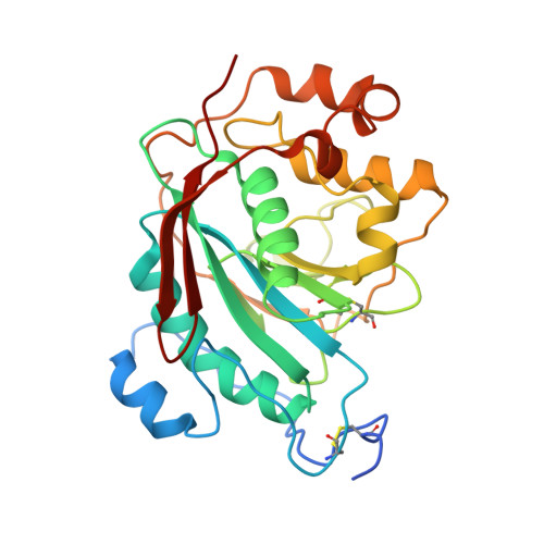
    6-sulfate modified N-acetylglucosamine (6-sulfo-GlcNAc) is often found as part of many biologically important carbohydrate epitopes such as 6-sulfo-Le(X). In these epitopes, the 6-sulfo-GlcNAc moiety is extended by a galactose sugar in a β1-4 linkage. The β4GalT1 enzyme transfers galactose (Gal) from UDP-Gal to N-acetylglucosamine (GlcNAc) in the presence of manganese. Here we report that the β4GalT1 enzyme transfers Gal to the 6-sulfo-GlcNAc and 4-methylumbelliferyl-6-sulfo-N-acetyl-β-D-glucosaminide (6-sulfo-βGlcNAc-MU) acceptor substrates, although with very low efficiency. To understand the effect that the 6-sulfate group on the GlcNAc acceptor has on the catalytic activity of the β4GalT1 molecule, we have determined the crystal structure of the catalytic domain of bovine β4GalT1 mutant enzyme M344H-β4GalT1 complex with the 6-sulfo-GlcNAc molecule. In the crystal structure, the 6-sulfo-GlcNAc is bound to the protein in a way that is similar to the GlcNAc molecule. However, the 6-sulfate group engages in additional interactions with the hydrophobic region, residues 276-285, of the protein molecule, and this group is found wedged between the aromatic side chains of Phe-280 and Trp314 residues. Since the side chain of the Trp314 residue undergoes conformational changes during the catalytic cycle of the enzyme, molecular interaction between Trp314 and the 6-sulfate group might hinder this conformational change. Therefore, the lack of a favorable binding environment, together with hindrance to the conformational changes, might be responsible for the poor catalytic activity.


  • Organizational Affiliation

    Structural Glycobiology Section, Basic Science Laboratory, National Cancer Institute at Frederick, P.O. Box B, Frederick, MD, 21702, USA.


Macromolecules
Find similar proteins by:  (by identity cutoff)  |  3D Structure
Entity ID: 1
MoleculeChains Sequence LengthOrganismDetailsImage
Beta-1,4-galactosyltransferase 1
A, B
286Bos taurusMutation(s): 2 
Gene Names: B4GALT1GALTGGTB2
EC: 2.4.1.22
UniProt
Find proteins for P08037 (Bos taurus)
Explore P08037 
Go to UniProtKB:  P08037
Entity Groups  
Sequence Clusters30% Identity50% Identity70% Identity90% Identity95% Identity100% Identity
UniProt GroupP08037
Sequence Annotations
Expand
  • Reference Sequence
Small Molecules
Ligands 4 Unique
IDChains Name / Formula / InChI Key2D Diagram3D Interactions
UDH
Query on UDH

Download Ideal Coordinates CCD File 
D [auth A],
M [auth B]
6-AMINOHEXYL-URIDINE-C1,5'-DIPHOSPHATE
C15 H27 N3 O12 P2
MLWJBKPFDKRHBM-FMKGYKFTSA-N
NGS
Query on NGS

Download Ideal Coordinates CCD File 
E [auth A],
N [auth B]
2-acetamido-2-deoxy-6-O-sulfo-beta-D-glucopyranose
C8 H15 N O9 S
WJFVEEAIYIOATH-FMDGEEDCSA-N
SO4
Query on SO4

Download Ideal Coordinates CCD File 
F [auth A]
G [auth A]
H [auth A]
I [auth A]
J [auth A]
F [auth A],
G [auth A],
H [auth A],
I [auth A],
J [auth A],
K [auth A],
O [auth B],
P [auth B],
Q [auth B],
R [auth B]
SULFATE ION
O4 S
QAOWNCQODCNURD-UHFFFAOYSA-L
MN
Query on MN

Download Ideal Coordinates CCD File 
C [auth A],
L [auth B]
MANGANESE (II) ION
Mn
WAEMQWOKJMHJLA-UHFFFAOYSA-N
Experimental Data & Validation

Experimental Data

  • Method: X-RAY DIFFRACTION
  • Resolution: 2.40 Å
  • R-Value Free: 0.249 
  • R-Value Work: 0.188 
  • R-Value Observed: 0.191 
  • Space Group: P 21 21 21
Unit Cell:
Length ( Å )Angle ( ˚ )
a = 49.282α = 90
b = 88.903β = 90
c = 139.734γ = 90
Software Package:
Software NamePurpose
PHENIXrefinement
REFMACrefinement
HKL-2000data collection
HKL-2000data reduction
HKL-2000data scaling
REFMACphasing

Structure Validation

View Full Validation Report


Ligand Structure Quality Assessment 


Entry History 

Deposition Data

Revision History  (Full details and data files)

  • Version 1.0: 2013-08-28
    Type: Initial release
  • Version 1.1: 2013-11-20
    Changes: Database references
  • Version 1.2: 2019-07-17
    Changes: Data collection, Refinement description
  • Version 2.0: 2020-07-29
    Type: Remediation
    Reason: Carbohydrate remediation
    Changes: Atomic model, Data collection, Database references, Derived calculations, Structure summary
  • Version 2.1: 2023-09-20
    Changes: Data collection, Database references, Refinement description, Structure summary