The role of the DNA-binding domains in CooA activation.
Kuchinskas, M., Li, H., Conrad, M., Roberts, G., Poulos, T.L.(2006) Biochemistry 45: 7148-7153
- PubMed: 16752905
- DOI: https://doi.org/10.1021/bi052609o
- Primary Citation of Related Structures:
4K8F - PubMed Abstract:
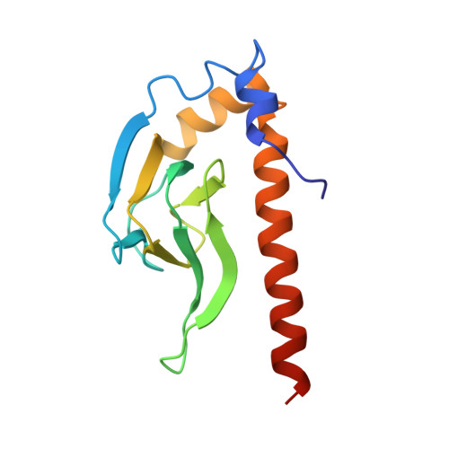
Carbon monoxide oxidation activator protein (CooA) is a dimeric carbon monoxide (CO) binding transcription factor that in the presence of CO promotes the transcription of genes involved in CO oxidation in Rhodospirillum rubrum. The off state (inactive) of Fe(II) CooA has His and Pro as the two axial heme ligands. In contrast, in the on state, which is active in DNA binding, the Pro ligand bond has been replaced by CO. This occurs by the transient loss of the Pro ligand, thus generating a pentacoordinate heme that can bind CO. The active on state of CooA has two major structural differences from the off state, in addition to the displacement of the Pro ligand by CO. There is a repositioning of long C helices at the dimer interface and a concomitant reorientation of the hinge region between the DNA- and effector-binding domains within each monomer [Roberts et al. (2005) J. Inorg. Biochem. 99, 280-292]. To better understand the relationship of these conformational changes, we have removed the DNA-binding domains and compared CO binding to the truncated and full-length protein. The crystal structure of truncated Fe(II) CooA is the same as that of the effector-binding domain of full-length Fe(II) CooA, including the relative orientation of the homodimer and the ligation environment of the heme. Thus, removing the DNA-binding domains has little obvious effect on the structure of CooA in the inactive off state. However, CO binds about 2-fold more tightly and about 10 times more rapidly to truncated CooA. The rate of CO binding is known to be limited by the dissociation of the Pro heme ligand [Puranik et al. (2004) J. Biol. Chem. 279, 21096-21108]. Therefore, the absence of the DNA-binding domain makes it easier for the Pro ligand to dissociate from the heme iron, which also makes it easier for truncated CooA to adopt the on-state structure.
Organizational Affiliation:
Departments of Molecular Biology and Biochemistry, University of California-Irvine, Irvine, California 92697-3990, USA.