Structural basis for recognition of synaptic vesicle protein 2C by botulinum neurotoxin A.
Benoit, R.M., Frey, D., Hilbert, M., Kevenaar, J.T., Wieser, M.M., Stirnimann, C.U., McMillan, D., Ceska, T., Lebon, F., Jaussi, R., Steinmetz, M.O., Schertler, G.F., Hoogenraad, C.C., Capitani, G., Kammerer, R.A.(2014) Nature 505: 108-111
- PubMed: 24240280
- DOI: https://doi.org/10.1038/nature12732
- Primary Citation of Related Structures:
4JRA - PubMed Abstract:
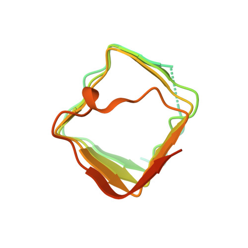
Botulinum neurotoxin A (BoNT/A) belongs to the most dangerous class of bioweapons. Despite this, BoNT/A is used to treat a wide range of common medical conditions such as migraines and a variety of ocular motility and movement disorders. BoNT/A is probably best known for its use as an antiwrinkle agent in cosmetic applications (including Botox and Dysport). BoNT/A application causes long-lasting flaccid paralysis of muscles through inhibiting the release of the neurotransmitter acetylcholine by cleaving synaptosomal-associated protein 25 (SNAP-25) within presynaptic nerve terminals. Two types of BoNT/A receptor have been identified, both of which are required for BoNT/A toxicity and are therefore likely to cooperate with each other: gangliosides and members of the synaptic vesicle glycoprotein 2 (SV2) family, which are putative transporter proteins that are predicted to have 12 transmembrane domains, associate with the receptor-binding domain of the toxin. Recently, fibroblast growth factor receptor 3 (FGFR3) has also been reported to be a potential BoNT/A receptor. In SV2 proteins, the BoNT/A-binding site has been mapped to the luminal domain, but the molecular details of the interaction between BoNT/A and SV2 are unknown. Here we determined the high-resolution crystal structure of the BoNT/A receptor-binding domain (BoNT/A-RBD) in complex with the SV2C luminal domain (SV2C-LD). SV2C-LD consists of a right-handed, quadrilateral β-helix that associates with BoNT/A-RBD mainly through backbone-to-backbone interactions at open β-strand edges, in a manner that resembles the inter-strand interactions in amyloid structures. Competition experiments identified a peptide that inhibits the formation of the complex. Our findings provide a strong platform for the development of novel antitoxin agents and for the rational design of BoNT/A variants with improved therapeutic properties.
Organizational Affiliation:
Laboratory of Biomolecular Research, Paul Scherrer Institut, CH-5232 Villigen PSI, Switzerland.