4JGF

Crystal Structure of the Cataract-Causing P23T gamma D-Crystallin Mutant


Experimental Data Snapshot

  • Method: X-RAY DIFFRACTION
  • Resolution: 2.50 Å
  • R-Value Free: 0.284 
  • R-Value Work: 0.228 
  • R-Value Observed: 0.233 

wwPDB Validation   3D Report Full Report


This is version 1.2 of the entry. See complete history


Literature

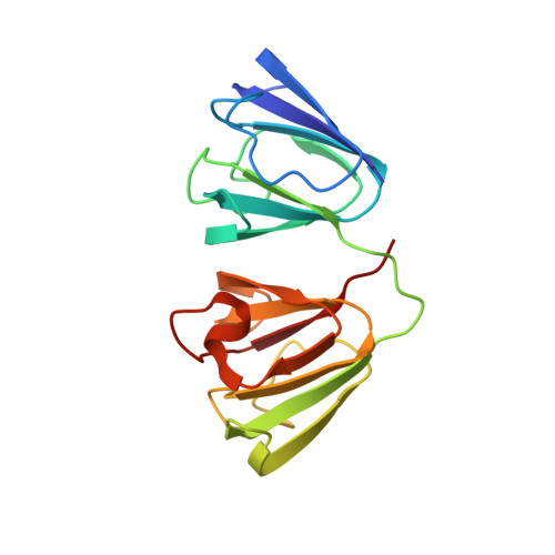
Crystal structure of the cataract-causing P23T gamma D-crystallin mutant.

Ji, F.Koharudin, L.M.Jung, J.Gronenborn, A.M.

(2013) Proteins 81: 1493-1498

  • DOI: https://doi.org/10.1002/prot.24321
  • Primary Citation of Related Structures:  
    4JGF

  • PubMed Abstract: 

    Up to now, efforts to crystallize the cataract-associated P23T mutant of human γD-crystallin have not been successful. Therefore, insights into the light scattering mechanism of this mutant have been exclusively obtained from solution work. Here we present the first crystal structure of the P23T mutant at 2.5 Å resolution. The protein exhibits essentially the same overall structure as seen for the wild-type protein. Based on our structural data, we confirm that no major conformational changes are caused by the mutation, and that solution phase properties of the mutant appear exclusively associated with cataract formation.


  • Organizational Affiliation

    Department of Structural Biology, University of Pittsburgh School of Medicine, Pittsburgh, Pennsylvania, 15261; School of Life Science and Biotechnology, Dalian University of Technology, Lingong Road, Dalian, 16024, China.


Macromolecules
Find similar proteins by:  (by identity cutoff)  |  3D Structure
Entity ID: 1
MoleculeChains Sequence LengthOrganismDetailsImage
Gamma-crystallin D
A, B
171Homo sapiensMutation(s): 1 
Gene Names: CRYGDCRYG4
UniProt & NIH Common Fund Data Resources
Find proteins for P07320 (Homo sapiens)
Explore P07320 
Go to UniProtKB:  P07320
PHAROS:  P07320
GTEx:  ENSG00000118231 
Entity Groups  
Sequence Clusters30% Identity50% Identity70% Identity90% Identity95% Identity100% Identity
UniProt GroupP07320
Sequence Annotations
Expand
  • Reference Sequence
Experimental Data & Validation

Experimental Data

  • Method: X-RAY DIFFRACTION
  • Resolution: 2.50 Å
  • R-Value Free: 0.284 
  • R-Value Work: 0.228 
  • R-Value Observed: 0.233 
  • Space Group: P 1 21 1
Unit Cell:
Length ( Å )Angle ( ˚ )
a = 37.55α = 90
b = 101.931β = 96.54
c = 41.371γ = 90
Software Package:
Software NamePurpose
StructureStudiodata collection
PHASERphasing
REFMACrefinement
d*TREKdata reduction
d*TREKdata scaling

Structure Validation

View Full Validation Report


Entry History 

Deposition Data

Revision History  (Full details and data files)

  • Version 1.0: 2013-05-22
    Type: Initial release
  • Version 1.1: 2013-10-02
    Changes: Database references
  • Version 1.2: 2024-02-28
    Changes: Data collection, Database references