


Crystal structure of human dihydroorotate dehydrogenase (DHODH) with DH03A016
Zhu, L., Zhu, J., Ren, X., Li, H.To be published.
Experimental Data Snapshot
Entity ID: 1 | |||||
|---|---|---|---|---|---|
| Molecule | Chains | Sequence Length | Organism | Details | Image |
| Dihydroorotate dehydrogenase (quinone), mitochondrial | 390 | Homo sapiens | Mutation(s): 0 Gene Names: DHODH EC: 1.3.5.2 | 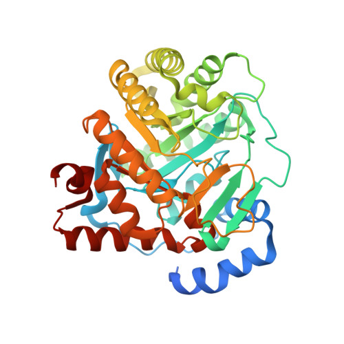 | |
UniProt & NIH Common Fund Data Resources | |||||
Find proteins for Q02127 (Homo sapiens) Explore Q02127 Go to UniProtKB: Q02127 | |||||
PHAROS: Q02127 GTEx: ENSG00000102967 | |||||
Entity Groups | |||||
| Sequence Clusters | 30% Identity50% Identity70% Identity90% Identity95% Identity100% Identity | ||||
| UniProt Group | Q02127 | ||||
Sequence AnnotationsExpand | |||||
| |||||
| Ligands 5 Unique | |||||
|---|---|---|---|---|---|
| ID | Chains | Name / Formula / InChI Key | 2D Diagram | 3D Interactions | |
| FMN Query on FMN | B [auth A] | FLAVIN MONONUCLEOTIDE C17 H21 N4 O9 P FVTCRASFADXXNN-SCRDCRAPSA-N |  | ||
| 4JG Query on 4JG | N [auth A] | 1-[4-methyl-2-(naphthalen-2-ylamino)-1,3-thiazol-5-yl]ethanone C16 H14 N2 O S CELUMPGHYUOWHA-UHFFFAOYSA-N |  | ||
| DET Query on DET | D [auth A] | UNDECYLAMINE-N,N-DIMETHYL-N-OXIDE C13 H29 N O OZHBUVQCJMARBN-UHFFFAOYSA-N |  | ||
| ORO Query on ORO | C [auth A] | OROTIC ACID C5 H4 N2 O4 PXQPEWDEAKTCGB-UHFFFAOYSA-N |  | ||
| SO4 Query on SO4 | E [auth A] F [auth A] G [auth A] H [auth A] I [auth A] | SULFATE ION O4 S QAOWNCQODCNURD-UHFFFAOYSA-L |  | ||
| Length ( Å ) | Angle ( ˚ ) |
|---|---|
| a = 90.76 | α = 90 |
| b = 90.76 | β = 90 |
| c = 123.49 | γ = 120 |
| Software Name | Purpose |
|---|---|
| HKL-2000 | data collection |
| PHASES | phasing |
| PHENIX | refinement |
| HKL-2000 | data reduction |
| HKL-2000 | data scaling |
| REFMAC | refinement |
RCSB PDB (citation) is hosted by
RCSB PDB is a member of the

