4HWT

Crystal structure of human Threonyl-tRNA synthetase bound to a novel inhibitor


Experimental Data Snapshot

  • Method: X-RAY DIFFRACTION
  • Resolution: 2.30 Å
  • R-Value Free: 0.257 
  • R-Value Work: 0.207 
  • R-Value Observed: 0.210 

wwPDB Validation   3D Report Full Report


Ligand Structure Quality Assessment 


This is version 1.0 of the entry. See complete history


Literature

Identification of bacteria-selective threonyl-tRNA synthetase substrate inhibitors by structure-based design.

Teng, M.Hilgers, M.T.Cunningham, M.L.Borchardt, A.Locke, J.B.Abraham, S.Haley, G.Kwan, B.P.Hall, C.Hough, G.W.Shaw, K.J.Finn, J.

(2013) J Med Chem 56: 1748-1760

  • DOI: https://doi.org/10.1021/jm301756m
  • Primary Citation of Related Structures:  
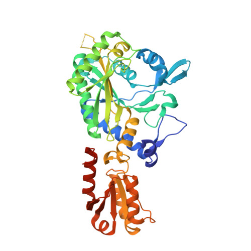
    4HWO, 4HWP, 4HWR, 4HWS, 4HWT

  • PubMed Abstract: 

    A series of potent and bacteria-selective threonyl-tRNA synthetase (ThrRS) inhibitors have been identified using structure-based drug design. These compounds occupied the substrate binding site of ThrRS and showed excellent binding affinities for all of the bacterial orthologues tested. Some of the compounds displayed greatly improved bacterial selectivity. Key residues responsible for potency and bacteria/human ThrRS selectivity have been identified. Antimicrobial activity has been achieved against wild-type Haemophilus influenzae and efflux-deficient mutants of Escherichia coli and Burkholderia thailandensis.


  • Organizational Affiliation

    Medicinal Chemistry, Trius Therapeutics, Inc., 6310 Nancy Ridge Drive, San Diego, California 92121, United States. mteng@triusrx.com


Macromolecules
Find similar proteins by:  (by identity cutoff)  |  3D Structure
Entity ID: 1
MoleculeChains Sequence LengthOrganismDetailsImage
Threonine--tRNA ligase, cytoplasmic
A, B
413Homo sapiensMutation(s): 0 
Gene Names: TARS
EC: 6.1.1.3
UniProt & NIH Common Fund Data Resources
Find proteins for P26639 (Homo sapiens)
Explore P26639 
Go to UniProtKB:  P26639
PHAROS:  P26639
GTEx:  ENSG00000113407 
Entity Groups  
Sequence Clusters30% Identity50% Identity70% Identity90% Identity95% Identity100% Identity
UniProt GroupP26639
Sequence Annotations
Expand
  • Reference Sequence
Small Molecules
Binding Affinity Annotations 
IDSourceBinding Affinity
1B2 Binding MOAD:  4HWT Ki: 5.00e+4 (nM) from 1 assay(s)
PDBBind:  4HWT Ki: 5.00e+4 (nM) from 1 assay(s)
Experimental Data & Validation

Experimental Data

  • Method: X-RAY DIFFRACTION
  • Resolution: 2.30 Å
  • R-Value Free: 0.257 
  • R-Value Work: 0.207 
  • R-Value Observed: 0.210 
  • Space Group: P 41 21 2
Unit Cell:
Length ( Å )Angle ( ˚ )
a = 73.897α = 90
b = 73.897β = 90
c = 357.587γ = 90
Software Package:
Software NamePurpose
d*TREKdata reduction
PHASERphasing
REFMACrefinement
PDB_EXTRACTdata extraction
d*TREKdata scaling

Structure Validation

View Full Validation Report


Ligand Structure Quality Assessment 


Entry History 

Deposition Data

Revision History  (Full details and data files)

  • Version 1.0: 2013-09-18
    Type: Initial release