4GV8

DUTPase from phage phi11 of S.aureus: visualization of the species-specific insert


Experimental Data Snapshot

  • Method: X-RAY DIFFRACTION
  • Resolution: 2.10 Å
  • R-Value Free: 0.232 
  • R-Value Work: 0.183 
  • R-Value Observed: 0.186 

wwPDB Validation   3D Report Full Report


Ligand Structure Quality Assessment 


This is version 1.3 of the entry. See complete history


Literature

Structure and enzymatic mechanism of a moonlighting dUTPase

Leveles, I.Nemeth, V.Szabo, J.E.Harmat, V.Nyiri, K.Bendes, A.A.Papp-Kadar, V.Zagyva, I.Rona, G.Ozohanics, O.Vekey, K.Toth, J.Vertessy, B.G.

(2013) Acta Crystallogr D Biol Crystallogr 69: 2298-2308

  • DOI: https://doi.org/10.1107/S0907444913021136
  • Primary Citation of Related Structures:  
    4GV8

  • PubMed Abstract: 

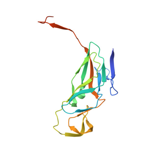
    Genome integrity requires well controlled cellular pools of nucleotides. dUTPases are responsible for regulating cellular dUTP levels and providing dUMP for dTTP biosynthesis. In Staphylococcus, phage dUTPases are also suggested to be involved in a moonlighting function regulating the expression of pathogenicity-island genes. Staphylococcal phage trimeric dUTPase sequences include a specific insertion that is not found in other organisms. Here, a 2.1 Å resolution three-dimensional structure of a ϕ11 phage dUTPase trimer with complete localization of the phage-specific insert, which folds into a small β-pleated mini-domain reaching out from the dUTPase core surface, is presented. The insert mini-domains jointly coordinate a single Mg2+ ion per trimer at the entrance to the threefold inner channel. Structural results provide an explanation for the role of Asp95, which is suggested to have functional significance in the moonlighting activity, as the metal-ion-coordinating moiety potentially involved in correct positioning of the insert. Enzyme-kinetics studies of wild-type and mutant constructs show that the insert has no major role in dUTP binding or cleavage and provide a description of the elementary steps (fast binding of substrate and release of products). In conclusion, the structural and kinetic data allow insights into both the phage-specific characteristics and the generally conserved traits of ϕ11 phage dUTPase.


  • Organizational Affiliation

    Institute of Enzymology, Research Centre for Natural Sciences, Hungarian Academy of Sciences, 29 Karolina Street, 1113 Budapest, Hungary.


Macromolecules
Find similar proteins by:  (by identity cutoff)  |  3D Structure
Entity ID: 1
MoleculeChains Sequence LengthOrganismDetailsImage
DUTPase
A, B, C, D, E
A, B, C, D, E, F
169Dubowvirus dv11Mutation(s): 0 
EC: 3.6.1.23
UniProt
Find proteins for Q8SDV3 (Staphylococcus phage phi 11)
Explore Q8SDV3 
Go to UniProtKB:  Q8SDV3
Entity Groups  
Sequence Clusters30% Identity50% Identity70% Identity90% Identity95% Identity100% Identity
UniProt GroupQ8SDV3
Sequence Annotations
Expand
  • Reference Sequence
Small Molecules
Ligands 2 Unique
IDChains Name / Formula / InChI Key2D Diagram3D Interactions
DUP
Query on DUP

Download Ideal Coordinates CCD File 
G [auth A]
J [auth B]
L [auth C]
N [auth D]
Q [auth E]
G [auth A],
J [auth B],
L [auth C],
N [auth D],
Q [auth E],
S [auth F]
2'-DEOXYURIDINE 5'-ALPHA,BETA-IMIDO-TRIPHOSPHATE
C9 H16 N3 O13 P3
XZLLMTSKYYYJLH-SHYZEUOFSA-N
MG
Query on MG

Download Ideal Coordinates CCD File 
H [auth A]
I [auth A]
K [auth B]
M [auth C]
O [auth D]
H [auth A],
I [auth A],
K [auth B],
M [auth C],
O [auth D],
P [auth D],
R [auth E],
T [auth F]
MAGNESIUM ION
Mg
JLVVSXFLKOJNIY-UHFFFAOYSA-N
Binding Affinity Annotations 
IDSourceBinding Affinity
DUP Binding MOAD:  4GV8 Kd: 320 (nM) from 1 assay(s)
PDBBind:  4GV8 Kd: 320 (nM) from 1 assay(s)
Experimental Data & Validation

Experimental Data

  • Method: X-RAY DIFFRACTION
  • Resolution: 2.10 Å
  • R-Value Free: 0.232 
  • R-Value Work: 0.183 
  • R-Value Observed: 0.186 
  • Space Group: P 43 2 2
Unit Cell:
Length ( Å )Angle ( ˚ )
a = 109.66α = 90
b = 109.66β = 90
c = 169.27γ = 90
Software Package:
Software NamePurpose
MOLREPphasing
REFMACrefinement
XDSdata reduction
XSCALEdata scaling

Structure Validation

View Full Validation Report


Ligand Structure Quality Assessment 


Entry History 

Deposition Data

Revision History  (Full details and data files)

  • Version 1.0: 2013-09-11
    Type: Initial release
  • Version 1.1: 2014-01-01
    Changes: Database references
  • Version 1.2: 2017-11-15
    Changes: Refinement description
  • Version 1.3: 2023-11-08
    Changes: Data collection, Database references, Derived calculations, Refinement description