4FN0

Crystal structure of mouse nectin-2 extracellular fragment D1-D2, 2nd crystal form


Experimental Data Snapshot

  • Method: X-RAY DIFFRACTION
  • Resolution: 3.35 Å
  • R-Value Free: 0.293 
  • R-Value Work: 0.247 
  • R-Value Observed: 0.249 

wwPDB Validation   3D Report Full Report


Ligand Structure Quality Assessment 


This is version 2.1 of the entry. See complete history


Literature

Nectin ectodomain structures reveal a canonical adhesive interface.

Harrison, O.J.Vendome, J.Brasch, J.Jin, X.Hong, S.Katsamba, P.S.Ahlsen, G.Troyanovsky, R.B.Troyanovsky, S.M.Honig, B.Shapiro, L.

(2012) Nat Struct Mol Biol 19: 906-915

  • DOI: https://doi.org/10.1038/nsmb.2366
  • Primary Citation of Related Structures:  
    4FMF, 4FMK, 4FN0, 4FOM, 4FQP, 4FRW, 4FS0

  • PubMed Abstract: 

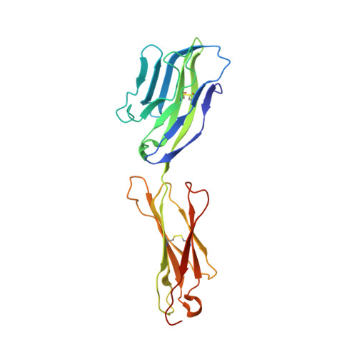
    Nectins are immunoglobulin superfamily glycoproteins that mediate intercellular adhesion in many vertebrate tissues. Homophilic and heterophilic interactions between nectin family members help mediate tissue patterning. We determined the homophilic binding affinities and heterophilic specificities of all four nectins and the related protein nectin-like 5 (Necl-5) from human and mouse, revealing a range of homophilic interaction strengths and a defined heterophilic specificity pattern. To understand the molecular basis of their adhesion and specificity, we determined the crystal structures of natively glycosylated full ectodomains or adhesive fragments of all four nectins and Necl-5. All of the crystal structures revealed dimeric nectins bound through a stereotyped interface that was previously proposed to represent a cis dimer. However, conservation of this interface and the results of targeted cross-linking experiments showed that this dimer probably represents the adhesive trans interaction. The structure of the dimer provides a simple molecular explanation for the adhesive binding specificity of nectins.


  • Organizational Affiliation

    Department of Biochemistry and Molecular Biophysics, Columbia University, New York, New York, USA.


Macromolecules
Find similar proteins by:  (by identity cutoff)  |  3D Structure
Entity ID: 1
MoleculeChains Sequence LengthOrganismDetailsImage
Poliovirus receptor-related protein 2
A, B, C
225Mus musculusMutation(s): 0 
Gene Names: Pvrl2MphPvrPvs
UniProt
Find proteins for P32507 (Mus musculus)
Explore P32507 
Go to UniProtKB:  P32507
Entity Groups  
Sequence Clusters30% Identity50% Identity70% Identity90% Identity95% Identity100% Identity
UniProt GroupP32507
Sequence Annotations
Expand
  • Reference Sequence
Oligosaccharides

Help

Entity ID: 2
MoleculeChains Length2D Diagram Glycosylation3D Interactions
beta-D-mannopyranose-(1-4)-2-acetamido-2-deoxy-beta-D-glucopyranose-(1-4)-[alpha-L-fucopyranose-(1-6)]2-acetamido-2-deoxy-beta-D-glucopyranose
D, E, F, G, H
4N-Glycosylation
Glycosylation Resources
GlyTouCan:  G32152BH
GlyCosmos:  G32152BH
GlyGen:  G32152BH
Small Molecules
Ligands 1 Unique
IDChains Name / Formula / InChI Key2D Diagram3D Interactions
NAG
Query on NAG

Download Ideal Coordinates CCD File 
I [auth C]2-acetamido-2-deoxy-beta-D-glucopyranose
C8 H15 N O6
OVRNDRQMDRJTHS-FMDGEEDCSA-N
Experimental Data & Validation

Experimental Data

  • Method: X-RAY DIFFRACTION
  • Resolution: 3.35 Å
  • R-Value Free: 0.293 
  • R-Value Work: 0.247 
  • R-Value Observed: 0.249 
  • Space Group: P 31 2 1
Unit Cell:
Length ( Å )Angle ( ˚ )
a = 117.022α = 90
b = 117.022β = 90
c = 158.467γ = 120
Software Package:
Software NamePurpose
ADSCdata collection
PHASERphasing
REFMACrefinement
HKL-2000data reduction
HKL-2000data scaling

Structure Validation

View Full Validation Report


Ligand Structure Quality Assessment 


Entry History 

Deposition Data

Revision History  (Full details and data files)

  • Version 1.0: 2012-08-22
    Type: Initial release
  • Version 1.1: 2012-09-05
    Changes: Database references
  • Version 1.2: 2012-09-26
    Changes: Database references
  • Version 2.0: 2020-07-29
    Type: Remediation
    Reason: Carbohydrate remediation
    Changes: Advisory, Atomic model, Data collection, Database references, Derived calculations, Structure summary
  • Version 2.1: 2023-09-13
    Changes: Data collection, Database references, Refinement description, Structure summary