4E7S

Myosin VI D23R I24R R569E (MD) pre-powerstroke state


Experimental Data Snapshot

  • Method: X-RAY DIFFRACTION
  • Resolution: 2.25 Å
  • R-Value Free: 0.216 
  • R-Value Work: 0.179 
  • R-Value Observed: 0.181 

wwPDB Validation   3D Report Full Report


Ligand Structure Quality Assessment 


This is version 1.3 of the entry. See complete history


Literature

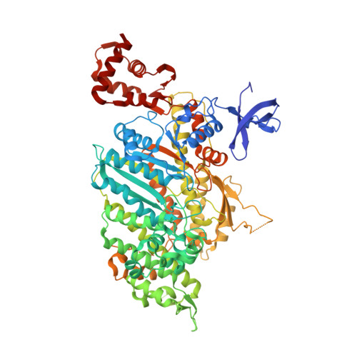
Processive Steps in the Reverse Direction Require Uncoupling of the Lead Head Lever Arm of Myosin VI.

Menetrey, J.Isabet, T.Ropars, V.Mukherjea, M.Pylypenko, O.Liu, X.Perez, J.Vachette, P.Sweeney, H.L.Houdusse, A.M.

(2012) Mol Cell 48: 75-86

  • DOI: https://doi.org/10.1016/j.molcel.2012.07.034
  • Primary Citation of Related Structures:  
    4ANJ, 4E7S, 4E7Z

  • PubMed Abstract: 

    Myosin VI is the only known reverse-direction myosin motor. It has an unprecedented means of amplifying movements within the motor involving rearrangements of the converter subdomain at the C terminus of the motor and an unusual lever arm projecting from the converter. While the average step size of a myosin VI dimer is 30-36 nm, the step size is highly variable, presenting a challenge to the lever arm mechanism by which all myosins are thought to move. Herein, we present structures of myosin VI that reveal regions of compliance that allow an uncoupling of the lead head when movement is modeled on actin. The location of the compliance restricts the possible actin binding sites and predicts the observed stepping behavior. The model reveals that myosin VI, unlike plus-end directed myosins, does not use a pure lever arm mechanism, but instead steps with a mechanism analogous to the kinesin neck-linker uncoupling model.


  • Organizational Affiliation

    Structural Motility, Institut Curie, Centre de Recherche, Paris, France.


Macromolecules
Find similar proteins by:  (by identity cutoff)  |  3D Structure
Entity ID: 1
MoleculeChains Sequence LengthOrganismDetailsImage
Myosin-VI
A, B
798Sus scrofaMutation(s): 3 
Gene Names: MYO6
UniProt
Find proteins for F1RQI7 (Sus scrofa)
Explore F1RQI7 
Go to UniProtKB:  F1RQI7
Entity Groups  
Sequence Clusters30% Identity50% Identity70% Identity90% Identity95% Identity100% Identity
UniProt GroupF1RQI7
Sequence Annotations
Expand
  • Reference Sequence
Small Molecules
Ligands 5 Unique
IDChains Name / Formula / InChI Key2D Diagram3D Interactions
ADP
Query on ADP

Download Ideal Coordinates CCD File 
C [auth A],
J [auth B]
ADENOSINE-5'-DIPHOSPHATE
C10 H15 N5 O10 P2
XTWYTFMLZFPYCI-KQYNXXCUSA-N
VO4
Query on VO4

Download Ideal Coordinates CCD File 
D [auth A],
K [auth B]
VANADATE ION
O4 V
LSGOVYNHVSXFFJ-UHFFFAOYSA-N
PO4
Query on PO4

Download Ideal Coordinates CCD File 
M [auth B]PHOSPHATE ION
O4 P
NBIIXXVUZAFLBC-UHFFFAOYSA-K
GOL
Query on GOL

Download Ideal Coordinates CCD File 
F [auth A]
G [auth A]
H [auth A]
I [auth A]
N [auth B]
F [auth A],
G [auth A],
H [auth A],
I [auth A],
N [auth B],
O [auth B]
GLYCEROL
C3 H8 O3
PEDCQBHIVMGVHV-UHFFFAOYSA-N
MG
Query on MG

Download Ideal Coordinates CCD File 
E [auth A],
L [auth B]
MAGNESIUM ION
Mg
JLVVSXFLKOJNIY-UHFFFAOYSA-N
Experimental Data & Validation

Experimental Data

  • Method: X-RAY DIFFRACTION
  • Resolution: 2.25 Å
  • R-Value Free: 0.216 
  • R-Value Work: 0.179 
  • R-Value Observed: 0.181 
  • Space Group: P 21 21 21
Unit Cell:
Length ( Å )Angle ( ˚ )
a = 81.947α = 90
b = 135.14β = 90
c = 196.54γ = 90
Software Package:
Software NamePurpose
ADSCdata collection
PHASERphasing
PHENIXrefinement
XDSdata reduction
XSCALEdata scaling

Structure Validation

View Full Validation Report


Ligand Structure Quality Assessment 


Entry History 

Deposition Data

Revision History  (Full details and data files)

  • Version 1.0: 2012-09-19
    Type: Initial release
  • Version 1.1: 2012-10-31
    Changes: Database references
  • Version 1.2: 2020-02-26
    Changes: Data collection, Database references
  • Version 1.3: 2024-02-28
    Changes: Data collection, Database references, Derived calculations