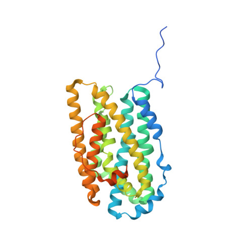
The Dimanganese(II) Site of Bacillus subtilis Class Ib Ribonucleotide Reductase.
Boal, A.K., Cotruvo, J.A., Stubbe, J., Rosenzweig, A.C.(2012) Biochemistry 51: 3861-3871
- PubMed: 22443445
- DOI: https://doi.org/10.1021/bi201925t
- Primary Citation of Related Structures:
4DR0 - PubMed Abstract:
Class Ib ribonucleotide reductases (RNRs) use a dimanganese-tyrosyl radical cofactor, Mn(III)(2)-Y(•), in their homodimeric NrdF (β2) subunit to initiate reduction of ribonucleotides to deoxyribonucleotides. The structure of the Mn(II)(2) form of NrdF is an important component in understanding O(2)-mediated formation of the active metallocofactor, a subject of much interest because a unique flavodoxin, NrdI, is required for cofactor assembly. Biochemical studies and sequence alignments suggest that NrdF and NrdI proteins diverge into three phylogenetically distinct groups. The only crystal structure to date of a NrdF with a fully ordered and occupied dimanganese site is that of Escherichia coli Mn(II)(2)-NrdF, prototypical of the enzymes from actinobacteria and proteobacteria. Here we report the 1.9 Å resolution crystal structure of Bacillus subtilis Mn(II)(2)-NrdF, representative of the enzymes from a second group, from Bacillus and Staphylococcus. The structures of the metal clusters in the β2 dimer are distinct from those observed in E. coli Mn(II)(2)-NrdF. These differences illustrate the key role that solvent molecules and protein residues in the second coordination sphere of the Mn(II)(2) cluster play in determining conformations of carboxylate residues at the metal sites and demonstrate that diverse coordination geometries are capable of serving as starting points for Mn(III)(2)-Y(•) cofactor assembly in class Ib RNRs.
Organizational Affiliation:
Department of Molecular Biosciences, Northwestern University, Evanston, Illinois 60208, USA.