Structure of the SthK Carboxy-Terminal Region Reveals a Gating Mechanism for Cyclic Nucleotide-Modulated Ion Channels.
Kesters, D., Brams, M., Nys, M., Wijckmans, E., Spurny, R., Voets, T., Tytgat, J., Kusch, J., Ulens, C.(2015) PLoS One 10: 16369
- PubMed: 25625648
- DOI: https://doi.org/10.1371/journal.pone.0116369
- Primary Citation of Related Structures:
4D7S, 4D7T - PubMed Abstract:
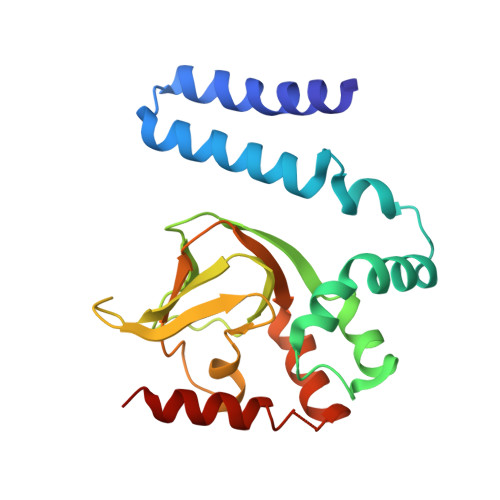
Cyclic nucleotide-sensitive ion channels are molecular pores that open in response to cAMP or cGMP, which are universal second messengers. Binding of a cyclic nucleotide to the carboxyterminal cyclic nucleotide binding domain (CNBD) of these channels is thought to cause a conformational change that promotes channel opening. The C-linker domain, which connects the channel pore to this CNBD, plays an important role in coupling ligand binding to channel opening. Current structural insight into this mechanism mainly derives from X-ray crystal structures of the C-linker/CNBD from hyperpolarization-activated cyclic nucleotide-modulated (HCN) channels. However, these structures reveal little to no conformational changes upon comparison of the ligand-bound and unbound form. In this study, we take advantage of a recently identified prokaryote ion channel, SthK, which has functional properties that strongly resemble cyclic nucleotide-gated (CNG) channels and is activated by cAMP, but not by cGMP. We determined X-ray crystal structures of the C-linker/CNBD of SthK in the presence of cAMP or cGMP. We observe that the structure in complex with cGMP, which is an antagonist, is similar to previously determined HCN channel structures. In contrast, the structure in complex with cAMP, which is an agonist, is in a more open conformation. We observe that the CNBD makes an outward swinging movement, which is accompanied by an opening of the C-linker. This conformation mirrors the open gate structures of the Kv1.2 channel or MthK channel, which suggests that the cAMP-bound C-linker/CNBD from SthK represents an activated conformation. These results provide a structural framework for better understanding cyclic nucleotide modulation of ion channels, including HCN and CNG channels.
Organizational Affiliation:
Laboratory of Structural Neurobiology, KU Leuven, Herestraat 49, PB601, Leuven, B-3000, Belgium.


