4CYB

DpsC from Streptomyces coelicolor


Experimental Data Snapshot

  • Method: X-RAY DIFFRACTION
  • Resolution: 1.78 Å
  • R-Value Free: 0.194 
  • R-Value Work: 0.159 
  • R-Value Observed: 0.161 

wwPDB Validation   3D Report Full Report


This is version 1.1 of the entry. See complete history


Literature

A Tale of Tails: Deciphering the Contribution of Terminal Tails to the Biochemical Properties of Two Dps Proteins from Streptomyces Coelicolor

Hitchings, M.D.Townsend, P.D.Pohl, E.Facey, P.D.Jones, D.H.Dyson, P.J.Del Sol, R.

(2014) Cell Mol Life Sci 71: 4911

  • DOI: https://doi.org/10.1007/s00018-014-1658-4
  • Primary Citation of Related Structures:  
    4CY9, 4CYA, 4CYB

  • PubMed Abstract: 

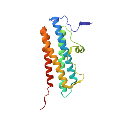
    Dps proteins are members of an extensive family of proteins that oxidise and deposit iron in the form of ferric oxide, and are also able to bind DNA. Ferroxidation centres are formed at the interface of anti-parallel dimers, which further assemble into dodecameric nanocages with a hollow core where ferric oxide is deposited. Streptomyces coelicolor encodes three Dps-like proteins (DpsA, B and C). Despite sharing the conserved four-helix bundle organisation observed in members of the Dps family, they display significant differences in the length of terminal extensions, or tails. DpsA possess both N- and C-terminal tails of different lengths, and their removal affects quaternary structure assembly to varying degrees. DpsC quaternary structure, on the other hand, is heavily dependent on its N-terminal tail as its removal abolishes correct protein folding. Analysis of the crystal structure of dodecamers from both proteins revealed remarkable differences in the position of tails and interface surface area; and provides insight to explain the differences in biochemical behaviour observed while comparing DpsA and DpsC.


  • Organizational Affiliation

    College of Medicine, Swansea University, Singleton Park, Swansea, SA2 8PP, UK.


Macromolecules
Find similar proteins by:  (by identity cutoff)  |  3D Structure
Entity ID: 1
MoleculeChains Sequence LengthOrganismDetailsImage
PUTATIVE DNA PROTECTION PROTEIN
A, B, C, D, E
A, B, C, D, E, F, G, H, I, J, K, L
173Streptomyces coelicolorMutation(s): 0 
UniProt
Find proteins for Q9K3L0 (Streptomyces coelicolor (strain ATCC BAA-471 / A3(2) / M145))
Explore Q9K3L0 
Go to UniProtKB:  Q9K3L0
Entity Groups  
Sequence Clusters30% Identity50% Identity70% Identity90% Identity95% Identity100% Identity
UniProt GroupQ9K3L0
Sequence Annotations
Expand
  • Reference Sequence
Small Molecules
Ligands 2 Unique
IDChains Name / Formula / InChI Key2D Diagram3D Interactions
FE
Query on FE

Download Ideal Coordinates CCD File 
AA [auth L]
M [auth A]
N [auth B]
O [auth C]
Q [auth D]
AA [auth L],
M [auth A],
N [auth B],
O [auth C],
Q [auth D],
R [auth E],
S [auth F],
T [auth G],
U [auth H],
V [auth I],
W [auth J],
Y [auth K]
FE (III) ION
Fe
VTLYFUHAOXGGBS-UHFFFAOYSA-N
NA
Query on NA

Download Ideal Coordinates CCD File 
BA [auth L],
P [auth C],
X [auth J],
Z [auth K]
SODIUM ION
Na
FKNQFGJONOIPTF-UHFFFAOYSA-N
Experimental Data & Validation

Experimental Data

  • Method: X-RAY DIFFRACTION
  • Resolution: 1.78 Å
  • R-Value Free: 0.194 
  • R-Value Work: 0.159 
  • R-Value Observed: 0.161 
  • Space Group: P 21 21 21
Unit Cell:
Length ( Å )Angle ( ˚ )
a = 92.22α = 90
b = 153.12β = 90
c = 170.28γ = 90
Software Package:
Software NamePurpose
REFMACrefinement
PHASERphasing

Structure Validation

View Full Validation Report


Entry History 

Deposition Data

Revision History  (Full details and data files)

  • Version 1.0: 2014-06-25
    Type: Initial release
  • Version 1.1: 2014-11-26
    Changes: Database references