4CQK

Crystal structure of ligand-bound NaD1


Experimental Data Snapshot

  • Method: X-RAY DIFFRACTION
  • Resolution: 1.60 Å
  • R-Value Free: 0.183 
  • R-Value Work: 0.155 
  • R-Value Observed: 0.156 

wwPDB Validation   3D Report Full Report


Ligand Structure Quality Assessment 


This is version 1.1 of the entry. See complete history


Literature

Phosphoinositide-Mediated Oligomerization of a Defensin Induces Cell Lysis.

Poon, I.K.H.Baxter, A.A.Lay, F.T.Mills, G.D.Adda, C.G.Payne, J.A.Phan, T.K.Ryan, G.F.White, J.A.Veneer, P.K.Van Der Weerden, N.L.Anderson, M.A.Kvansakul, M.Hulett, M.D.

(2014) Elife 3: 1808

  • DOI: https://doi.org/10.7554/eLife.01808
  • Primary Citation of Related Structures:  
    4CQK

  • PubMed Abstract: 

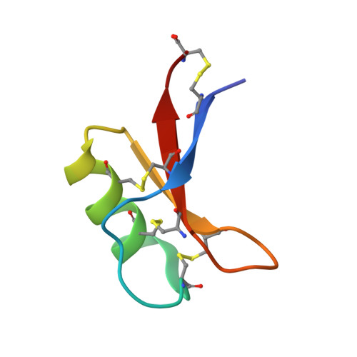
    Cationic antimicrobial peptides (CAPs) such as defensins are ubiquitously found innate immune molecules that often exhibit broad activity against microbial pathogens and mammalian tumor cells. Many CAPs act at the plasma membrane of cells leading to membrane destabilization and permeabilization. In this study, we describe a novel cell lysis mechanism for fungal and tumor cells by the plant defensin NaD1 that acts via direct binding to the plasma membrane phospholipid phosphatidylinositol 4,5-bisphosphate (PIP2). We determined the crystal structure of a NaD1:PIP2 complex, revealing a striking oligomeric arrangement comprising seven dimers of NaD1 that cooperatively bind the anionic headgroups of 14 PIP2 molecules through a unique 'cationic grip' configuration. Site-directed mutagenesis of NaD1 confirms that PIP2-mediated oligomerization is important for fungal and tumor cell permeabilization. These observations identify an innate recognition system by NaD1 for direct binding of PIP2 that permeabilizes cells via a novel membrane disrupting mechanism. DOI: http://dx.doi.org/10.7554/eLife.01808.001.


  • Organizational Affiliation

    Department of Biochemistry, La Trobe Institute for Molecular Science, La Trobe University, Melbourne, Australia.


Macromolecules
Find similar proteins by:  (by identity cutoff)  |  3D Structure
Entity ID: 1
MoleculeChains Sequence LengthOrganismDetailsImage
FLOWER-SPECIFIC DEFENSIN
A, B, C, D, E
A, B, C, D, E, F, G, H, I, J, K, L, M, N
47Nicotiana alataMutation(s): 0 
Membrane Entity: Yes 
UniProt
Find proteins for Q8GTM0 (Nicotiana alata)
Explore Q8GTM0 
Go to UniProtKB:  Q8GTM0
Entity Groups  
Sequence Clusters30% Identity50% Identity70% Identity90% Identity95% Identity100% Identity
UniProt GroupQ8GTM0
Sequence Annotations
Expand
  • Reference Sequence
Small Molecules
Ligands 3 Unique
IDChains Name / Formula / InChI Key2D Diagram3D Interactions
PIO
Query on PIO

Download Ideal Coordinates CCD File 
DA [auth F]
GA [auth G]
JA [auth H]
LA [auth I]
NA [auth J]
DA [auth F],
GA [auth G],
JA [auth H],
LA [auth I],
NA [auth J],
O [auth A],
PA [auth K],
RA [auth L],
S [auth B],
T [auth C],
UA [auth M],
W [auth D],
XA [auth N],
Y [auth E]
[(2R)-2-octanoyloxy-3-[oxidanyl-[(1R,2R,3S,4R,5R,6S)-2,3,6-tris(oxidanyl)-4,5-diphosphonooxy-cyclohexyl]oxy-phosphoryl]oxy-propyl] octanoate
C25 H49 O19 P3
XLNCEHRXXWQMPK-MJUMVPIBSA-N
MRD
Query on MRD

Download Ideal Coordinates CCD File 
BA [auth E],
CA [auth E],
FA [auth F],
R [auth A]
(4R)-2-METHYLPENTANE-2,4-DIOL
C6 H14 O2
SVTBMSDMJJWYQN-RXMQYKEDSA-N
SO4
Query on SO4

Download Ideal Coordinates CCD File 
AA [auth E]
EA [auth F]
HA [auth G]
IA [auth G]
KA [auth H]
AA [auth E],
EA [auth F],
HA [auth G],
IA [auth G],
KA [auth H],
MA [auth I],
OA [auth J],
P [auth A],
Q [auth A],
QA [auth K],
SA [auth L],
TA [auth L],
U [auth C],
V [auth C],
VA [auth M],
WA [auth M],
X [auth D],
YA [auth N],
Z [auth E],
ZA [auth N]
SULFATE ION
O4 S
QAOWNCQODCNURD-UHFFFAOYSA-L
Experimental Data & Validation

Experimental Data

Unit Cell:
Length ( Å )Angle ( ˚ )
a = 79.64α = 90
b = 132.04β = 90
c = 153.01γ = 90
Software Package:
Software NamePurpose
PHENIXrefinement
XDSdata reduction
SCALAdata scaling
PHASERphasing

Structure Validation

View Full Validation Report


Ligand Structure Quality Assessment 


Entry History 

Deposition Data

Revision History  (Full details and data files)

  • Version 1.0: 2014-04-16
    Type: Initial release
  • Version 1.1: 2023-12-20
    Changes: Data collection, Database references, Derived calculations, Other, Refinement description