4CMY

Chlorobium tepidum Ferritin


Experimental Data Snapshot

  • Method: X-RAY DIFFRACTION
  • Resolution: 2.59 Å
  • R-Value Free: 0.218 
  • R-Value Work: 0.193 
  • R-Value Observed: 0.194 

wwPDB Validation   3D Report Full Report


This is version 1.2 of the entry. See complete history


Literature

The Crystal Structure of Ferritin from Chlorobium Tepidum Reveals a New Conformation of the 4-Fold Channel for This Protein Family.

Arenas, M.Townsend, P.D.Brito, C.Marquez, V.Marabolli, V.Gonzalez-Nilo, F.Matias, C.Watt, R.K.Lopez-Castro, J.D.Dominguez-Vera, J.Pohl, E.Yevenes, A.

(2014) Biochimie 106: 39

  • DOI: https://doi.org/10.1016/j.biochi.2014.07.019
  • Primary Citation of Related Structures:  
    4CMY

  • PubMed Abstract: 

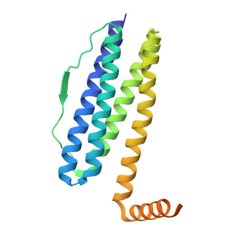
    Ferritins are ubiquitous iron-storage proteins found in all kingdoms of life. They share a common architecture made of 24 subunits of five α-helices. The recombinant Chlorobium tepidum ferritin (rCtFtn) is a structurally interesting protein since sequence alignments with other ferritins show that this protein has a significantly extended C-terminus, which possesses 12 histidine residues as well as several aspartate and glutamic acid residues that are potential metal ion binding residues. We show that the macromolecular assembly of rCtFtn exhibits a cage-like hollow shell consisting of 24 monomers that are related by 4-3-2 symmetry; similar to the assembly of other ferritins. In all ferritins of known structure the short fifth α-helix adopts an acute angle with respect to the four-helix bundle. However, the crystal structure of the rCtFtn presented here shows that this helix adopts a new conformation defining a new assembly of the 4-fold channel of rCtFtn. This conformation allows the arrangement of the C-terminal region into the inner cavity of the protein shell. Furthermore, two Fe(III) ions were found in each ferroxidase center of rCtFtn, with an average FeA-FeB distance of 3 Å; corresponding to a diferric μ-oxo/hydroxo species. This is the first ferritin crystal structure with an isolated di-iron center in an iron-storage ferritin. The crystal structure of rCtFtn and the biochemical results presented here, suggests that rCtFtn presents similar biochemical properties reported for other members of this protein family albeit with distinct structural plasticity.


  • Organizational Affiliation

    Centro de Bioinformática y Simulación Molecular, Facultad de Ingeniería, Universidad de Talca, Talca, Chile.


Macromolecules
Find similar proteins by:  (by identity cutoff)  |  3D Structure
Entity ID: 1
MoleculeChains Sequence LengthOrganismDetailsImage
FERRITIN
A, B, C, D, E
A, B, C, D, E, F, G, H, I, J, K, L, M, N, O, P, Q, R, S, T, U, W, X
203Chlorobaculum tepidumMutation(s): 0 
UniProt
Find proteins for Q8KBP5 (Chlorobaculum tepidum (strain ATCC 49652 / DSM 12025 / NBRC 103806 / TLS))
Explore Q8KBP5 
Go to UniProtKB:  Q8KBP5
Entity Groups  
Sequence Clusters30% Identity50% Identity70% Identity90% Identity95% Identity100% Identity
UniProt GroupQ8KBP5
Sequence Annotations
Expand
  • Reference Sequence
Find similar proteins by:  (by identity cutoff)  |  3D Structure
Entity ID: 2
MoleculeChains Sequence LengthOrganismDetailsImage
FERRITIN203Chlorobaculum tepidumMutation(s): 0 
UniProt
Find proteins for Q8KBP5 (Chlorobaculum tepidum (strain ATCC 49652 / DSM 12025 / NBRC 103806 / TLS))
Explore Q8KBP5 
Go to UniProtKB:  Q8KBP5
Entity Groups  
Sequence Clusters30% Identity50% Identity70% Identity90% Identity95% Identity100% Identity
UniProt GroupQ8KBP5
Sequence Annotations
Expand
  • Reference Sequence
Small Molecules
Ligands 1 Unique
IDChains Name / Formula / InChI Key2D Diagram3D Interactions
FE
Query on FE

Download Ideal Coordinates CCD File 
AA [auth B]
AB [auth O]
BA [auth B]
BB [auth O]
CA [auth C]
AA [auth B],
AB [auth O],
BA [auth B],
BB [auth O],
CA [auth C],
CB [auth P],
DA [auth C],
DB [auth P],
EA [auth D],
EB [auth Q],
FA [auth D],
FB [auth Q],
GA [auth E],
GB [auth R],
HA [auth E],
HB [auth R],
IA [auth F],
IB [auth S],
JA [auth F],
JB [auth S],
KA [auth G],
KB [auth T],
LA [auth G],
LB [auth T],
MA [auth H],
MB [auth U],
NA [auth H],
NB [auth U],
OA [auth I],
OB [auth V],
PA [auth I],
PB [auth V],
QA [auth J],
QB [auth W],
RA [auth J],
RB [auth W],
SA [auth K],
SB [auth X],
TA [auth K],
TB [auth X],
UA [auth L],
VA [auth L],
WA [auth M],
XA [auth M],
Y [auth A],
YA [auth N],
Z [auth A],
ZA [auth N]
FE (III) ION
Fe
VTLYFUHAOXGGBS-UHFFFAOYSA-N
Experimental Data & Validation

Experimental Data

  • Method: X-RAY DIFFRACTION
  • Resolution: 2.59 Å
  • R-Value Free: 0.218 
  • R-Value Work: 0.193 
  • R-Value Observed: 0.194 
  • Space Group: P 21 21 21
Unit Cell:
Length ( Å )Angle ( ˚ )
a = 123.31α = 90
b = 190.37β = 90
c = 242.07γ = 90
Software Package:
Software NamePurpose
REFMACrefinement

Structure Validation

View Full Validation Report


Entry History 

Deposition Data

Revision History  (Full details and data files)

  • Version 1.0: 2014-11-26
    Type: Initial release
  • Version 1.1: 2014-12-03
    Changes: Database references
  • Version 1.2: 2015-08-12
    Changes: Database references