4APZ

Structure of B. subtilis genomic dUTPase YncF in complex with dU, PPi and Mg in P1


Experimental Data Snapshot

  • Method: X-RAY DIFFRACTION
  • Resolution: 2.01 Å
  • R-Value Free: 0.212 
  • R-Value Work: 0.169 
  • R-Value Observed: 0.171 

wwPDB Validation   3D Report Full Report


Ligand Structure Quality Assessment 


This is version 1.4 of the entry. See complete history


Literature

Tying Down the Arm in Bacillus Dutpase: Structure and Mechanism

Garcia-Nafria, J.Timm, J.Harrison, C.Turkenburg, J.P.Wilson, K.S.

(2013) Acta Crystallogr D Biol Crystallogr 69: 1367

  • DOI: https://doi.org/10.1107/S090744491300735X
  • Primary Citation of Related Structures:  
    4AO5, 4AOO, 4AOZ, 4APZ, 4B0H

  • PubMed Abstract: 

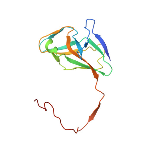
    Homotrimeric dUTPases contain three active sites, each formed by five conserved sequence motifs originating from all three subunits. The essential fifth motif lies in a flexible C-terminal arm which becomes ordered during catalysis and is disordered in most crystal structures. Previously, it has been shown that the two Bacillus subtilis dUTPases, YncF and YosS, differ from their orthologues in the position in the sequence of the essential Phe-lid residue, which stacks against the uracil base, and in the conformation of the general base aspartate, which points away from the active site. Here, three structures of the complex of YncF with dU-PPi-Mg(2+) and the structure of YosS complexed with dUMP are reported. dU-PPi-Mg(2+) triggers the ordering of both the C-terminal arm and a loop (residues 18-26) which is uniquely disordered in the Bacillus dUTPases. The dUMP complex suggests two stages in substrate release. Limited proteolysis experiments allowed those complexes in which C-terminal cleavage is hindered and those in which it can be assumed to be ordered to be identified. The results lead to the suggestion that dUpNHpp is not a perfect substrate mimic, at least for the B. subtilis enzymes, and provide new insights into the mechanism of these two dUTPases in comparison to their orthologues. The enzyme mechanism is reviewed using the present and previous crystal structures as snapshots along the reaction coordinate.


  • Organizational Affiliation

    Structural Biology Laboratory, Department of Chemistry, University of York, Heslington, York YO10 5DD, England.


Macromolecules
Find similar proteins by:  (by identity cutoff)  |  3D Structure
Entity ID: 1
MoleculeChains Sequence LengthOrganismDetailsImage
PROBABLE DEOXYURIDINE 5'-TRIPHOSPHATE NUCLEOTIDOHYDROLASE YNCF144Bacillus subtilisMutation(s): 0 
EC: 3.6.1.23
UniProt
Find proteins for O31801 (Bacillus subtilis (strain 168))
Explore O31801 
Go to UniProtKB:  O31801
Entity Groups  
Sequence Clusters30% Identity50% Identity70% Identity90% Identity95% Identity100% Identity
UniProt GroupO31801
Sequence Annotations
Expand
  • Reference Sequence
Small Molecules
Ligands 4 Unique
IDChains Name / Formula / InChI Key2D Diagram3D Interactions
DUR
Query on DUR

Download Ideal Coordinates CCD File 
AC [auth B]
AF [auth b]
BD [auth K]
CB [auth 3]
CE [auth T]
AC [auth B],
AF [auth b],
BD [auth K],
CB [auth 3],
CE [auth T],
CG [auth k],
DC [auth C],
DF [auth c],
ED [auth L],
FB [auth 4],
FE [auth U],
FG [auth l],
GC [auth D],
GF [auth d],
HD [auth M],
IB [auth 5],
IE [auth V],
JC [auth E],
JF [auth e],
JG [auth m],
KD [auth N],
LB [auth 6],
LE [auth W],
MC [auth F],
ND [auth O],
NF [auth f],
OB [auth 7],
OE [auth X],
PC [auth G],
QD [auth P],
QF [auth g],
RB [auth 8],
RE [auth Y],
SC [auth H],
TD [auth Q],
TF [auth h],
UB [auth 9],
UE [auth Z],
VC [auth I],
WA [auth 1],
WD [auth R],
WF [auth i],
XB [auth A],
XE [auth a],
YC [auth J],
ZA [auth 2],
ZD [auth S],
ZF [auth j]
2'-DEOXYURIDINE
C9 H12 N2 O5
MXHRCPNRJAMMIM-SHYZEUOFSA-N
POP
Query on POP

Download Ideal Coordinates CCD File 
AB [auth 2]
AE [auth S]
AG [auth j]
BC [auth B]
BF [auth b]
AB [auth 2],
AE [auth S],
AG [auth j],
BC [auth B],
BF [auth b],
CD [auth K],
DB [auth 3],
DE [auth T],
DG [auth k],
EC [auth C],
EF [auth c],
FD [auth L],
GB [auth 4],
GE [auth U],
GG [auth l],
HC [auth D],
HF [auth d],
ID [auth M],
JB [auth 5],
JE [auth V],
KC [auth E],
KF [auth e],
KG [auth m],
LD [auth N],
MB [auth 6],
ME [auth W],
NC [auth F],
OD [auth O],
OF [auth f],
PB [auth 7],
PE [auth X],
QC [auth G],
RD [auth P],
RF [auth g],
SB [auth 8],
SE [auth Y],
TC [auth H],
UD [auth Q],
UF [auth h],
VB [auth 9],
VE [auth Z],
WC [auth I],
XA [auth 1],
XD [auth R],
XF [auth i],
YB [auth A],
YE [auth a],
ZC [auth J]
PYROPHOSPHATE 2-
H2 O7 P2
XPPKVPWEQAFLFU-UHFFFAOYSA-L
PO4
Query on PO4

Download Ideal Coordinates CCD File 
IG [auth l],
MF [auth e]
PHOSPHATE ION
O4 P
NBIIXXVUZAFLBC-UHFFFAOYSA-K
MG
Query on MG

Download Ideal Coordinates CCD File 
AD [auth J]
BB [auth 2]
BE [auth S]
BG [auth j]
CC [auth B]
AD [auth J],
BB [auth 2],
BE [auth S],
BG [auth j],
CC [auth B],
CF [auth b],
DD [auth K],
EB [auth 3],
EE [auth T],
EG [auth k],
FC [auth C],
FF [auth c],
GD [auth L],
HB [auth 4],
HE [auth U],
HG [auth l],
IC [auth D],
IF [auth d],
JD [auth M],
KB [auth 5],
KE [auth V],
LC [auth E],
LF [auth e],
LG [auth m],
MD [auth N],
NB [auth 6],
NE [auth W],
OC [auth F],
PD [auth O],
PF [auth f],
QB [auth 7],
QE [auth X],
RC [auth G],
SD [auth P],
SF [auth g],
TB [auth 8],
TE [auth Y],
UC [auth H],
VD [auth Q],
VF [auth h],
WB [auth 9],
WE [auth Z],
XC [auth I],
YA [auth 1],
YD [auth R],
YF [auth i],
ZB [auth A],
ZE [auth a]
MAGNESIUM ION
Mg
JLVVSXFLKOJNIY-UHFFFAOYSA-N
Experimental Data & Validation

Experimental Data

  • Method: X-RAY DIFFRACTION
  • Resolution: 2.01 Å
  • R-Value Free: 0.212 
  • R-Value Work: 0.169 
  • R-Value Observed: 0.171 
  • Space Group: P 1
Unit Cell:
Length ( Å )Angle ( ˚ )
a = 97.06α = 89.72
b = 97.07β = 88.47
c = 193.85γ = 90.11
Software Package:
Software NamePurpose
REFMACrefinement
XDSdata reduction
SCALAdata scaling
MOLREPphasing

Structure Validation

View Full Validation Report


Ligand Structure Quality Assessment 


Entry History 

Deposition Data

Revision History  (Full details and data files)

  • Version 1.0: 2013-04-24
    Type: Initial release
  • Version 1.1: 2013-05-22
    Changes: Atomic model, Derived calculations, Non-polymer description, Other
  • Version 1.2: 2013-07-31
    Changes: Atomic model, Database references, Derived calculations, Other, Structure summary
  • Version 1.3: 2013-08-07
    Changes: Database references
  • Version 1.4: 2023-12-20
    Changes: Data collection, Database references, Derived calculations, Other, Refinement description