4ANP

Crystal structure of human phenylalanine hydroxylase in complex with a pharmacological chaperone


Experimental Data Snapshot

  • Method: X-RAY DIFFRACTION
  • Resolution: 2.11 Å
  • R-Value Free: 0.237 
  • R-Value Work: 0.204 
  • R-Value Observed: 0.205 

wwPDB Validation   3D Report Full Report


Ligand Structure Quality Assessment 


This is version 1.2 of the entry. See complete history


Literature

Structural and Mechanistic Basis of the Interaction between a Pharmacological Chaperone and Human Phenylalanine Hydroxylase.

Torreblanca, R.Lira-Navarrete, E.Sancho, J.Hurtado-Guerrero, R.

(2012) Chembiochem 13: 1266

  • DOI: https://doi.org/10.1002/cbic.201200188
  • Primary Citation of Related Structures:  
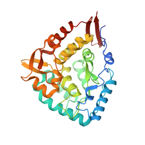
    4ANP

  • PubMed Abstract: 

    Not without a chaperone: Pharmacological chaperones are designed to bind and ideally stabilise their target protein. Here, we elucidate the molecular mechanism of a potential pharmacological chaperone to treat phenylketonuria. The crystal structure of human phenylalanine hydroxylase with compound IV may help in the rational design of more efficient compounds to treat this disease.


  • Organizational Affiliation

    Department of Biochemistry and Molecular and Cellular Biology, Institute of Biocomputation and Physics of Complex Systems (BIFI), University of Zaragoza, BIFI-IQFR (CSIC) Joint Unit, Pedro Cerbuna 12, Mariano Esquillor s/n, Campus Rio Ebro, Edificio I+D, Spain.


Macromolecules
Find similar proteins by:  (by identity cutoff)  |  3D Structure
Entity ID: 1
MoleculeChains Sequence LengthOrganismDetailsImage
PHENYLALANINE-4-HYDROXYLASE324Homo sapiensMutation(s): 0 
EC: 1.14.16.1
UniProt & NIH Common Fund Data Resources
Find proteins for P00439 (Homo sapiens)
Explore P00439 
Go to UniProtKB:  P00439
PHAROS:  P00439
GTEx:  ENSG00000171759 
Entity Groups  
Sequence Clusters30% Identity50% Identity70% Identity90% Identity95% Identity100% Identity
UniProt GroupP00439
Sequence Annotations
Expand
  • Reference Sequence
Small Molecules
Ligands 2 Unique
IDChains Name / Formula / InChI Key2D Diagram3D Interactions
3QI
Query on 3QI

Download Ideal Coordinates CCD File 
C [auth A]5,6-DIMETHYL-3-(4-METHYL-2-PYRIDINYL)-2-THIOXO-2,3-DIHYDROTHIENO[2,3- D]PYRIMIDIN-4(1H)-ONE
C14 H13 N3 O S2
NYDQMBWQPMNQIZ-UHFFFAOYSA-N
FE
Query on FE

Download Ideal Coordinates CCD File 
B [auth A]FE (III) ION
Fe
VTLYFUHAOXGGBS-UHFFFAOYSA-N
Binding Affinity Annotations 
IDSourceBinding Affinity
3QI Binding MOAD:  4ANP Ki: 2.00e+5 (nM) from 1 assay(s)
Experimental Data & Validation

Experimental Data

  • Method: X-RAY DIFFRACTION
  • Resolution: 2.11 Å
  • R-Value Free: 0.237 
  • R-Value Work: 0.204 
  • R-Value Observed: 0.205 
  • Space Group: C 2 2 21
Unit Cell:
Length ( Å )Angle ( ˚ )
a = 66.283α = 90
b = 107.511β = 90
c = 124.55γ = 90
Software Package:
Software NamePurpose
REFMACrefinement
SAINTdata reduction
SADABSdata scaling
MOLREPphasing

Structure Validation

View Full Validation Report


Ligand Structure Quality Assessment 


Entry History 

Deposition Data

Revision History  (Full details and data files)

  • Version 1.0: 2012-04-11
    Type: Initial release
  • Version 1.1: 2012-07-25
    Changes: Database references
  • Version 1.2: 2023-12-20
    Changes: Data collection, Database references, Derived calculations, Other, Refinement description