Structural Characterization of Bacterioferritin from Blastochloris Viridis.
Wahlgren, W.Y., Omran, H., von Stetten, D., Royant, A., van der Post, S., Katona, G.(2012) PLoS One 7: 46992
- PubMed: 23056552
- DOI: https://doi.org/10.1371/journal.pone.0046992
- Primary Citation of Related Structures:
4AM2, 4AM4, 4AM5 - PubMed Abstract:
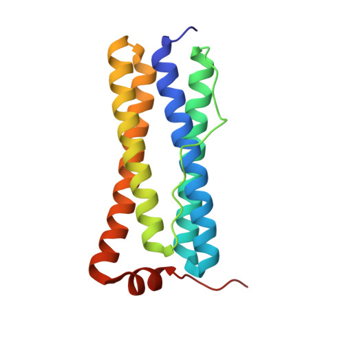
Iron storage and elimination of toxic ferrous iron are the responsibility of bacterioferritins in bacterial species. Bacterioferritins are capable of oxidizing iron using molecular oxygen and import iron ions into the large central cavity of the protein, where they are stored in a mineralized form. We isolated, crystallized bacterioferritin from the microaerophilic/anaerobic, purple non-sulfur bacterium Blastochloris viridis and determined its amino acid sequence and X-ray structure. The structure and sequence revealed similarity to other purple bacterial species with substantial differences in the pore regions. Static 3- and 4-fold pores do not allow the passage of iron ions even though structural dynamics may assist the iron gating. On the other hand the B-pore is open to water and larger ions in its native state. In order to study the mechanism of iron import, multiple soaking experiments were performed. Upon Fe(II) and urea treatment the ferroxidase site undergoes reorganization as seen in bacterioferritin from Escherichia coli and Pseudomonas aeruginosa. When soaking with Fe(II) only, a closely bound small molecular ligand is observed close to Fe(1) and the coordination of Glu94 to Fe(2) changes from bidentate to monodentate. DFT calculations indicate that the bound ligand is most likely a water or a hydroxide molecule representing a product complex. On the other hand the different soaking treatments did not modify the conformation of other pore regions.
Organizational Affiliation:
Department of Chemistry and Molecular Biology, University of Gothenburg, Göteborg, Sweden.