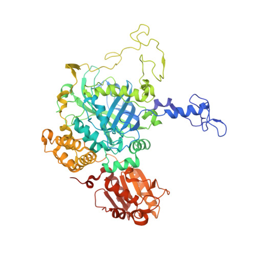
4AJ9

Catalase 3 from Neurospora crassa


Experimental Data Snapshot

  • Method: X-RAY DIFFRACTION
  • Resolution: 1.85 Å
  • R-Value Free: 0.218 
  • R-Value Work: 0.177 
  • R-Value Observed: 0.179 

wwPDB Validation   3D Report Full Report


Ligand Structure Quality Assessment 


This is version 1.2 of the entry. See complete history


Literature

X-ray driven reduction of Cpd I of Catalase-3 from N. crassa reveals differential sensitivity of active sites and formation of ferrous state.

Zarate-Romero, A.Stojanoff, V.Cohen, A.E.Hansberg, W.Rudino-Pinera, E.

(2019) Arch Biochem Biophys 666: 107-115

  • DOI: https://doi.org/10.1016/j.abb.2019.03.020
  • Primary Citation of Related Structures:  
    4AJ9, 6NSW, 6NSY, 6NSZ, 6NT0, 6NT1

  • PubMed Abstract: 

    Catalases are biotechnologically relevant enzymes because of their applications in food technology, bioremediation, and biomedicine. The dismutation of hydrogen peroxide occurs in two steps; in the first one, the enzyme forms an oxidized compound I (Cpd I) and in the second one, the enzyme is reduced to the ferric state. In this research work, we analyzed the reduction of Cpd I by X-ray radiation damage during diffraction experiments in crystals of CAT-3, a Large-Size Subunit Catalase (LSC) from Neurospora crassa. A Multi-Crystal Data collection Strategy was applied in order to obtain the Cpd I structure at a resolution of 2.2 Å; this intermediate was highly sensitive to X-ray and was easily reduced at very low deposited radiation dose, causing breakage of the Fe=O bond. The comparison of the structures showed reduced intermediates and also evidenced the differential sensitivity per monomer. The resting ferric state was reduced to the ferrous state, an intermediate without a previous report in LSC. The chemically obtained Cpd I and the X-ray reduced intermediates were identified by UV-visible microspectrometry coupled to data collection. The differential sensitivity and the formation of a ferrous state are discussed, emphasizing the importance of the correct interpretation in the oxidation state of the iron heme.


  • Organizational Affiliation

    Cátedras CONACyT, Departamento de Bionanotecnología, Centro de Nanociencias y Nanotecnología, Universidad Nacional Autónoma de México, Ensenada, B. C., Mexico.


Macromolecules
Find similar proteins by:  (by identity cutoff)  |  3D Structure
Entity ID: 1
MoleculeChains Sequence LengthOrganismDetailsImage
CATALASE-3
A, B, C, D
682Neurospora crassaMutation(s): 0 
EC: 1.11.1.6
UniProt
Find proteins for Q9C169 (Neurospora crassa (strain ATCC 24698 / 74-OR23-1A / CBS 708.71 / DSM 1257 / FGSC 987))
Explore Q9C169 
Go to UniProtKB:  Q9C169
Entity Groups  
Sequence Clusters30% Identity50% Identity70% Identity90% Identity95% Identity100% Identity
UniProt GroupQ9C169
Sequence Annotations
Expand
  • Reference Sequence
Small Molecules
Ligands 3 Unique
IDChains Name / Formula / InChI Key2D Diagram3D Interactions
HEM
Query on HEM

Download Ideal Coordinates CCD File 
E [auth A],
GA [auth D],
O [auth B],
Y [auth C]
PROTOPORPHYRIN IX CONTAINING FE
C34 H32 Fe N4 O4
KABFMIBPWCXCRK-RGGAHWMASA-L
1PE
Query on 1PE

Download Ideal Coordinates CCD File 
BA [auth C]
CA [auth C]
DA [auth C]
EA [auth C]
FA [auth C]
BA [auth C],
CA [auth C],
DA [auth C],
EA [auth C],
FA [auth C],
H [auth A],
I [auth A],
J [auth A],
JA [auth D],
K [auth A],
KA [auth D],
L [auth A],
LA [auth D],
M [auth A],
MA [auth D],
N [auth A],
NA [auth D],
R [auth B],
S [auth B],
T [auth B],
U [auth B],
V [auth B],
W [auth B],
X [auth B]
PENTAETHYLENE GLYCOL
C10 H22 O6
JLFNLZLINWHATN-UHFFFAOYSA-N
ACT
Query on ACT

Download Ideal Coordinates CCD File 
AA [auth C]
F [auth A]
G [auth A]
HA [auth D]
IA [auth D]
AA [auth C],
F [auth A],
G [auth A],
HA [auth D],
IA [auth D],
P [auth B],
Q [auth B],
Z [auth C]
ACETATE ION
C2 H3 O2
QTBSBXVTEAMEQO-UHFFFAOYSA-M
Experimental Data & Validation

Experimental Data

  • Method: X-RAY DIFFRACTION
  • Resolution: 1.85 Å
  • R-Value Free: 0.218 
  • R-Value Work: 0.177 
  • R-Value Observed: 0.179 
  • Space Group: P 21 21 21
Unit Cell:
Length ( Å )Angle ( ˚ )
a = 130.75α = 90
b = 154.19β = 90
c = 160.3γ = 90
Software Package:
Software NamePurpose
PHENIXrefinement
XDSdata reduction
XSCALEdata scaling
PHASERphasing

Structure Validation

View Full Validation Report


Ligand Structure Quality Assessment 


Entry History 

Deposition Data

Revision History  (Full details and data files)

  • Version 1.0: 2013-03-06
    Type: Initial release
  • Version 1.1: 2019-04-24
    Changes: Data collection, Database references, Other
  • Version 1.2: 2023-12-20
    Changes: Data collection, Database references, Derived calculations, Other, Refinement description