


Crystal Structure of Human P63 Tetramerization Domain
Muniz, J.R.C., Coutandin, D., Salah, E., Chaikuad, A., Vollmar, M., Weigelt, J., Arrowsmith, C.H., Edwards, A.M., Bountra, C., Dotsch, V., von Delft, F., Knapp, S.To be published.
Experimental Data Snapshot
wwPDB Validation 3D Report Full Report

Entity ID: 1 | |||||
|---|---|---|---|---|---|
| Molecule | Chains | Sequence Length | Organism | Details | Image |
| TUMOR PROTEIN 63 | 61 | Homo sapiens | Mutation(s): 0 | 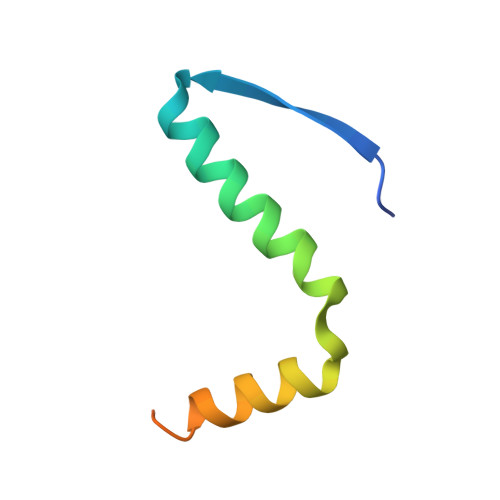 | |
UniProt & NIH Common Fund Data Resources | |||||
Find proteins for Q9H3D4 (Homo sapiens) Explore Q9H3D4 Go to UniProtKB: Q9H3D4 | |||||
PHAROS: Q9H3D4 GTEx: ENSG00000073282 | |||||
Entity Groups | |||||
| Sequence Clusters | 30% Identity50% Identity70% Identity90% Identity95% Identity100% Identity | ||||
| UniProt Group | Q9H3D4 | ||||
Sequence AnnotationsExpand | |||||
| |||||
| Ligands 1 Unique | |||||
|---|---|---|---|---|---|
| ID | Chains | Name / Formula / InChI Key | 2D Diagram | 3D Interactions | |
| PE4 Query on PE4 | E [auth A] | 2-{2-[2-(2-{2-[2-(2-ETHOXY-ETHOXY)-ETHOXY]-ETHOXY}-ETHOXY)-ETHOXY]-ETHOXY}-ETHANOL C16 H34 O8 PJWQOENWHPEPKI-UHFFFAOYSA-N |  | ||
| Length ( Å ) | Angle ( ˚ ) |
|---|---|
| a = 80.85 | α = 90 |
| b = 80.85 | β = 90 |
| c = 68.37 | γ = 120 |
| Software Name | Purpose |
|---|---|
| BUSTER | refinement |
| MOSFLM | data reduction |
| SCALA | data scaling |
| PHASER | phasing |
RCSB PDB (citation) is hosted by
RCSB PDB is a member of the

