4A6F

Crystal structure of Slm1-PH domain in complex with Phosphoserine


Experimental Data Snapshot

  • Method: X-RAY DIFFRACTION
  • Resolution: 1.68 Å
  • R-Value Free: 0.242 
  • R-Value Work: 0.214 
  • R-Value Observed: 0.215 

wwPDB Validation   3D Report Full Report


Ligand Structure Quality Assessment 


This is version 1.3 of the entry. See complete history


Literature

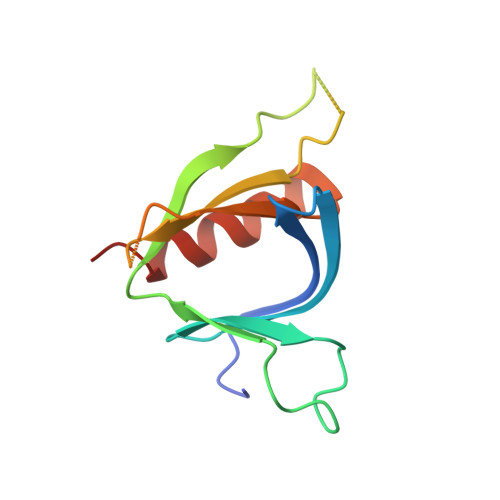
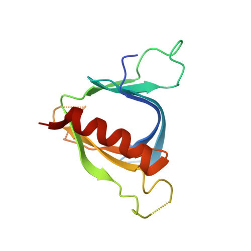
Structural Analyses of Slm1-Ph Domain Demonstrate Ligand Binding in the Non-Canonical Site

Anand, K.Maeda, K.Gavin, A.C.

(2012) PLoS One 7: 36526

  • DOI: https://doi.org/10.1371/journal.pone.0036526
  • Primary Citation of Related Structures:  
    4A5K, 4A6F, 4A6H, 4A6K

  • PubMed Abstract: 

    Pleckstrin homology (PH) domains are common membrane-targeting modules and their best characterized ligands are a set of important signaling lipids that include phosphatidylinositol phosphates (PtdInsPs). PH domains recognize PtdInsPs through two distinct mechanisms that use different binding pockets on opposite sides of the β-strands 1 and 2: i) a canonical binding site delimited by the β1-β2 and β3-β4loops and ii) a non-canonical binding site bordered by the β1-β2 and β5-β6loops. The PH domain-containing protein Slm1 from budding yeast Saccharomyces cerevisiae is required for actin cytoskeleton polarization and cell growth. We recently reported that this PH domain binds PtdInsPs and phosphorylated sphingolipids in a cooperative manner.


  • Organizational Affiliation

    European Molecular Biology Laboratory Heidelberg, Structural and Computational Biology Unit, Heidelberg, Germany. anand@embl.de


Macromolecules
Find similar proteins by:  (by identity cutoff)  |  3D Structure
Entity ID: 1
MoleculeChains Sequence LengthOrganismDetailsImage
PHOSPHATIDYLINOSITOL 4,5-BISPHOSPHATE-BINDING PROTEIN SLM1120Saccharomyces cerevisiaeMutation(s): 0 
Membrane Entity: Yes 
UniProt
Find proteins for P40485 (Saccharomyces cerevisiae (strain ATCC 204508 / S288c))
Explore P40485 
Go to UniProtKB:  P40485
Entity Groups  
Sequence Clusters30% Identity50% Identity70% Identity90% Identity95% Identity100% Identity
UniProt GroupP40485
Sequence Annotations
Expand
  • Reference Sequence
Find similar proteins by:  (by identity cutoff)  |  3D Structure
Entity ID: 2
MoleculeChains Sequence LengthOrganismDetailsImage
PHOSPHATIDYLINOSITOL 4,5-BISPHOSPHATE-BINDING PROTEIN SLM1120Saccharomyces cerevisiaeMutation(s): 0 
Membrane Entity: Yes 
UniProt
Find proteins for P40485 (Saccharomyces cerevisiae (strain ATCC 204508 / S288c))
Explore P40485 
Go to UniProtKB:  P40485
Entity Groups  
Sequence Clusters30% Identity50% Identity70% Identity90% Identity95% Identity100% Identity
UniProt GroupP40485
Sequence Annotations
Expand
  • Reference Sequence
Experimental Data & Validation

Experimental Data

  • Method: X-RAY DIFFRACTION
  • Resolution: 1.68 Å
  • R-Value Free: 0.242 
  • R-Value Work: 0.214 
  • R-Value Observed: 0.215 
  • Space Group: P 2 21 21
Unit Cell:
Length ( Å )Angle ( ˚ )
a = 37.54α = 90
b = 73.65β = 90
c = 82.47γ = 90
Software Package:
Software NamePurpose
PHENIXrefinement
XDSdata reduction
XSCALEdata scaling
PHASERphasing

Structure Validation

View Full Validation Report


Ligand Structure Quality Assessment 


Entry History 

Deposition Data

Revision History  (Full details and data files)

  • Version 1.0: 2012-06-13
    Type: Initial release
  • Version 1.1: 2013-02-06
    Changes: Atomic model
  • Version 1.2: 2019-10-16
    Changes: Data collection, Other
  • Version 1.3: 2023-12-20
    Changes: Data collection, Database references, Derived calculations, Refinement description