4A63

Crystal structure of the p73-ASPP2 complex at 2.6A resolution


Experimental Data Snapshot

  • Method: X-RAY DIFFRACTION
  • Resolution: 2.27 Å
  • R-Value Free: 0.244 
  • R-Value Work: 0.215 
  • R-Value Observed: 0.217 

wwPDB Validation   3D Report Full Report


This is version 1.4 of the entry. See complete history


Literature

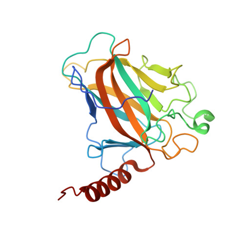
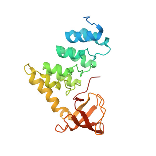
Structural Basis for Aspp2 Recognition by the Tumor Suppressor P73.

Canning, P.von Delft, F.Bullock, A.N.

(2012) J Mol Biol 423: 515

  • DOI: https://doi.org/10.1016/j.jmb.2012.08.005
  • Primary Citation of Related Structures:  
    2XWC, 4A63

  • PubMed Abstract: 

    Tumor suppressors p53, p63 and p73 comprise a family of stress-responsive transcription factors with distinct functions in development and tumor suppression. Most human cancers lose p53 function, yet all three proteins are capable of inducing apoptosis or cellular senescence. Mechanisms are therefore under investigation to activate p73-dependent apoptosis in p53-deficient cancer cells. Significantly, the DNA-binding domain (DBD) of p73 escapes viral oncoproteins and displays an enhanced thermal stability. To further understand the variant features of p73, we solved the high-resolution crystal structure of the p73 DBD as well as its complex with the ankyrin repeat and SH3 domains of the pro-apoptotic factor ASPP2. The p73 structure exhibits the same conserved architecture as p53 but displays a divergent L2 loop, a known site of protein-protein interaction. The loop in p73 is changed by a two-residue insertion that also induces repacking around the site of the p53 mutational hotspot R175. Importantly, the binding of ASPP2 is preserved by conformational changes in both the ankyrin repeat and SH3 domains. These results further highlight the structural variation that impacts p53 family interactions within the p53 interactome.


  • Organizational Affiliation

    Structural Genomics Consortium, University of Oxford, Old Road Campus, Roosevelt Drive, Oxford OX3 7DQ, UK.


Macromolecules
Find similar proteins by:  (by identity cutoff)  |  3D Structure
Entity ID: 1
MoleculeChains Sequence LengthOrganismDetailsImage
TUMOUR PROTEIN 73
A, C, E, G, I
A, C, E, G, I, K
208Homo sapiensMutation(s): 0 
UniProt & NIH Common Fund Data Resources
Find proteins for O15350 (Homo sapiens)
Explore O15350 
Go to UniProtKB:  O15350
PHAROS:  O15350
GTEx:  ENSG00000078900 
Entity Groups  
Sequence Clusters30% Identity50% Identity70% Identity90% Identity95% Identity100% Identity
UniProt GroupO15350
Sequence Annotations
Expand
  • Reference Sequence
Find similar proteins by:  (by identity cutoff)  |  3D Structure
Entity ID: 2
MoleculeChains Sequence LengthOrganismDetailsImage
APOPTOSIS STIMULATING OF P53 PROTEIN 2
B, D, F, H, J
B, D, F, H, J, L
239Homo sapiensMutation(s): 0 
UniProt & NIH Common Fund Data Resources
Find proteins for Q13625 (Homo sapiens)
Explore Q13625 
Go to UniProtKB:  Q13625
PHAROS:  Q13625
GTEx:  ENSG00000143514 
Entity Groups  
Sequence Clusters30% Identity50% Identity70% Identity90% Identity95% Identity100% Identity
UniProt GroupQ13625
Sequence Annotations
Expand
  • Reference Sequence
Small Molecules
Ligands 2 Unique
IDChains Name / Formula / InChI Key2D Diagram3D Interactions
ZN
Query on ZN

Download Ideal Coordinates CCD File 
M [auth C],
Q [auth E],
R [auth G],
T [auth I],
W [auth K]
ZINC ION
Zn
PTFCDOFLOPIGGS-UHFFFAOYSA-N
ACT
Query on ACT

Download Ideal Coordinates CCD File 
N [auth C]
O [auth C]
P [auth C]
S [auth G]
U [auth I]
N [auth C],
O [auth C],
P [auth C],
S [auth G],
U [auth I],
V [auth I],
X [auth K],
Y [auth K],
Z [auth L]
ACETATE ION
C2 H3 O2
QTBSBXVTEAMEQO-UHFFFAOYSA-M
Experimental Data & Validation

Experimental Data

  • Method: X-RAY DIFFRACTION
  • Resolution: 2.27 Å
  • R-Value Free: 0.244 
  • R-Value Work: 0.215 
  • R-Value Observed: 0.217 
  • Space Group: I 1 2 1
Unit Cell:
Length ( Å )Angle ( ˚ )
a = 132.81α = 90
b = 170.1β = 91.98
c = 177.555γ = 90
Software Package:
Software NamePurpose
BUSTERrefinement
MOSFLMdata reduction
SCALAdata scaling
PHASERphasing

Structure Validation

View Full Validation Report


Entry History 

Revision History  (Full details and data files)

  • Version 1.0: 2011-12-21
    Type: Initial release
  • Version 1.1: 2012-09-26
    Changes: Database references, Structure summary
  • Version 1.2: 2012-10-31
    Changes: Database references
  • Version 1.3: 2018-01-24
    Changes: Database references, Structure summary
  • Version 1.4: 2023-12-20
    Changes: Data collection, Database references, Derived calculations, Other, Refinement description