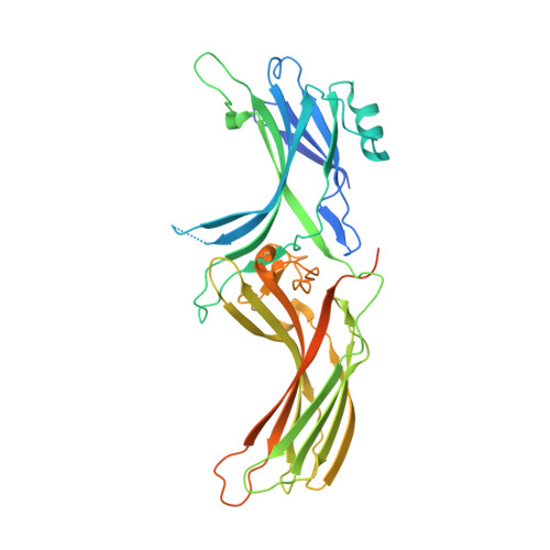
Structural evidence for the role of polar core residue Arg175 in arrestin activation.
Granzin, J., Stadler, A., Cousin, A., Schlesinger, R., Batra-Safferling, R.(2015) Sci Rep 5: 15808-15808
- PubMed: 26510463
- DOI: https://doi.org/10.1038/srep15808
- PubMed Abstract:
Binding mechanism of arrestin requires photoactivation and phosphorylation of the receptor protein rhodopsin, where the receptor bound phosphate groups cause displacement of the long C-tail 'activating' arrestin. Mutation of arginine 175 to glutamic acid (R175E), a central residue in the polar core and previously predicted as the 'phosphosensor' leads to a pre-active arrestin that is able to terminate phototransduction by binding to non-phosphorylated, light-activated rhodopsin. Here, we report the first crystal structure of a R175E mutant arrestin at 2.7 Å resolution that reveals significant differences compared to the basal state reported in full-length arrestin structures. These differences comprise disruption of hydrogen bond network in the polar core, and three-element interaction including disordering of several residues in the receptor-binding finger loop and the C-terminus (residues 361-404). Additionally, R175E structure shows a 7.5° rotation of the amino and carboxy-terminal domains relative to each other. Consistent to the biochemical data, our structure suggests an important role of R29 in the initial activation step of C-tail release. Comparison of the crystal structures of basal arrestin and R175E mutant provide insights into the mechanism of arrestin activation, where binding of the receptor likely induces structural changes mimicked as in R175E.
Organizational Affiliation:
Institute of Complex Systems (ICS-6), Structural Biochemistry, Forschungszentrum Jülich, 52425, Jülich, Germany.