4ZLP

Crystal Structure of Notch3 Negative Regulatory Region

  • Classification: TRANSCRIPTION
  • Organism(s): Homo sapiens
  • Expression System: Homo sapiens
  • Mutation(s): No 

  • Deposited: 2015-05-01 Released: 2015-08-19 
  • Deposition Author(s): Xu, X., Blacklow, S.C.
  • Funding Organization(s): National Institutes of Health/National Cancer Institute (NIH/NCI)

Experimental Data Snapshot

  • Method: X-RAY DIFFRACTION
  • Resolution: 2.48 Å
  • R-Value Free: 0.204 
  • R-Value Work: 0.178 
  • R-Value Observed: 0.179 

wwPDB Validation   3D Report Full Report


Ligand Structure Quality Assessment 


This is version 2.0 of the entry. See complete history


Literature

Insights into Autoregulation of Notch3 from Structural and Functional Studies of Its Negative Regulatory Region.

Xu, X.Choi, S.H.Hu, T.Tiyanont, K.Habets, R.Groot, A.J.Vooijs, M.Aster, J.C.Chopra, R.Fryer, C.Blacklow, S.C.

(2015) Structure 23: 1227-1235

  • DOI: https://doi.org/10.1016/j.str.2015.05.001
  • Primary Citation of Related Structures:  
    4ZLP

  • PubMed Abstract: 

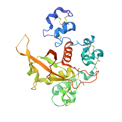
    Notch receptors are transmembrane proteins that undergo activating proteolysis in response to ligand stimulation. A negative regulatory region (NRR) maintains receptor quiescence by preventing protease cleavage prior to ligand binding. We report here the X-ray structure of the NRR of autoinhibited human Notch3, and compare it with the Notch1 and Notch2 NRRs. The overall architecture of the autoinhibited conformation, in which three LIN12-Notch repeat (LNR) modules wrap around a heterodimerization domain, is preserved in Notch3, but the autoinhibited conformation of the Notch3 NRR is less stable. The Notch3 NRR uses a highly conserved surface on the third LNR module to form a dimer in the crystal. Similar homotypic interfaces exist in Notch1 and Notch2. Together, these studies reveal distinguishing structural features associated with increased basal activity of Notch3, demonstrate increased ligand-independent signaling for disease-associated mutations that map to the Notch3 NRR, and identify a conserved dimerization interface present in multiple Notch receptors.


  • Organizational Affiliation

    Department of Cancer Biology, Dana Farber Cancer Institute, Boston, MA 02215, USA; Department of Biological Chemistry and Molecular Pharmacology, Harvard Medical School, Boston, MA 02115, USA.


Macromolecules
Find similar proteins by:  (by identity cutoff)  |  3D Structure
Entity ID: 1
MoleculeChains Sequence LengthOrganismDetailsImage
Neurogenic locus notch homolog protein 3
A, B
271Homo sapiensMutation(s): 0 
Gene Names: NOTCH3
UniProt & NIH Common Fund Data Resources
Find proteins for Q9UM47 (Homo sapiens)
Explore Q9UM47 
Go to UniProtKB:  Q9UM47
PHAROS:  Q9UM47
GTEx:  ENSG00000074181 
Entity Groups  
Sequence Clusters30% Identity50% Identity70% Identity90% Identity95% Identity100% Identity
UniProt GroupQ9UM47
Sequence Annotations
Expand
  • Reference Sequence
Oligosaccharides

Help

Entity ID: 2
MoleculeChains Length2D Diagram Glycosylation3D Interactions
alpha-D-mannopyranose-(1-6)-alpha-D-mannopyranose-(1-4)-2-acetamido-2-deoxy-beta-D-glucopyranose-(1-4)-2-acetamido-2-deoxy-beta-D-glucopyranose
C
4N-Glycosylation
Glycosylation Resources
GlyTouCan:  G04854NQ
GlyCosmos:  G04854NQ
GlyGen:  G04854NQ
Small Molecules
Ligands 4 Unique
IDChains Name / Formula / InChI Key2D Diagram3D Interactions
NAG
Query on NAG

Download Ideal Coordinates CCD File 
N [auth B]2-acetamido-2-deoxy-beta-D-glucopyranose
C8 H15 N O6
OVRNDRQMDRJTHS-FMDGEEDCSA-N
GOL
Query on GOL

Download Ideal Coordinates CCD File 
G [auth A],
H [auth A],
O [auth B]
GLYCEROL
C3 H8 O3
PEDCQBHIVMGVHV-UHFFFAOYSA-N
ACT
Query on ACT

Download Ideal Coordinates CCD File 
I [auth A],
J [auth A]
ACETATE ION
C2 H3 O2
QTBSBXVTEAMEQO-UHFFFAOYSA-M
CA
Query on CA

Download Ideal Coordinates CCD File 
D [auth A]
E [auth A]
F [auth A]
K [auth B]
L [auth B]
D [auth A],
E [auth A],
F [auth A],
K [auth B],
L [auth B],
M [auth B]
CALCIUM ION
Ca
BHPQYMZQTOCNFJ-UHFFFAOYSA-N
Experimental Data & Validation

Experimental Data

  • Method: X-RAY DIFFRACTION
  • Resolution: 2.48 Å
  • R-Value Free: 0.204 
  • R-Value Work: 0.178 
  • R-Value Observed: 0.179 
  • Space Group: C 1 2 1
Unit Cell:
Length ( Å )Angle ( ˚ )
a = 122.482α = 90
b = 64.31β = 105.23
c = 83.089γ = 90
Software Package:
Software NamePurpose
PHENIXrefinement
HKL-2000data reduction
HKL-2000data scaling
PHENIXphasing

Structure Validation

View Full Validation Report


Ligand Structure Quality Assessment 


Entry History & Funding Information

Deposition Data


Funding OrganizationLocationGrant Number
National Institutes of Health/National Cancer Institute (NIH/NCI)United StatesR01 CA092433 to Stephen C Blacklow

Revision History  (Full details and data files)

  • Version 1.0: 2015-08-19
    Type: Initial release
  • Version 1.1: 2017-09-20
    Changes: Author supporting evidence, Database references, Derived calculations
  • Version 1.2: 2019-12-04
    Changes: Author supporting evidence
  • Version 2.0: 2020-07-29
    Type: Remediation
    Reason: Carbohydrate remediation
    Changes: Advisory, Atomic model, Data collection, Derived calculations, Structure summary