4YPH

Crystal Structure of MutY bound to its anti-substrate with the disulfide cross-linker reduced


Experimental Data Snapshot

  • Method: X-RAY DIFFRACTION
  • Resolution: 2.32 Å
  • R-Value Free: 0.259 
  • R-Value Work: 0.187 
  • R-Value Observed: 0.191 

wwPDB Validation   3D Report Full Report


This is version 1.3 of the entry. See complete history


Literature

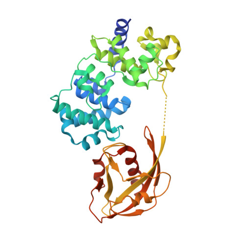
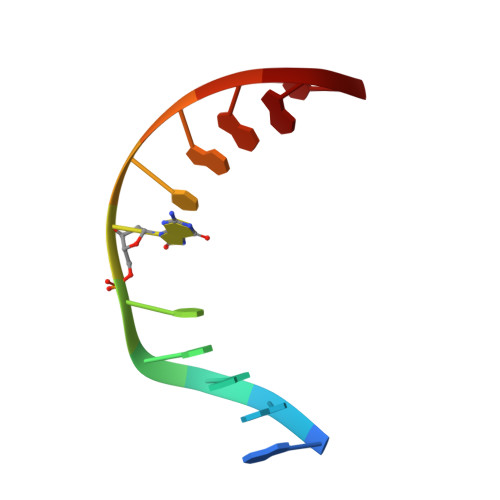
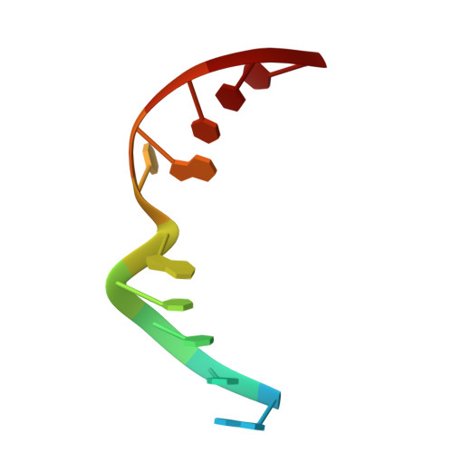
Structural Basis for Avoidance of Promutagenic DNA Repair by MutY Adenine DNA Glycosylase.

Wang, L.Lee, S.J.Verdine, G.L.

(2015) J Biol Chem 290: 17096-17105

  • DOI: https://doi.org/10.1074/jbc.M115.657866
  • Primary Citation of Related Structures:  
    4YOQ, 4YPH, 4YPR

  • PubMed Abstract: 

    The highly mutagenic A:oxoG (8-oxoguanine) base pair in DNA most frequently arises by aberrant replication of the primary oxidative lesion C:oxoG. This lesion is particularly insidious because neither of its constituent nucleobases faithfully transmit genetic information from the original C:G base pair. Repair of A:oxoG is initiated by adenine DNA glycosylase, which catalyzes hydrolytic cleavage of the aberrant A nucleobase from the DNA backbone. These enzymes, MutY in bacteria and MUTYH in humans, scrupulously avoid processing of C:oxoG because cleavage of the C residue in C:oxoG would actually promote mutagenic conversion to A:oxoG. Here we analyze the structural basis for rejection of C:oxoG by MutY, using a synthetic crystallography approach to capture the enzyme in the process of inspecting the C:oxoG anti-substrate, with which it ordinarily binds only fleetingly. We find that MutY uses two distinct strategies to avoid presentation of C to the enzyme active site. Firstly, MutY possesses an exo-site that serves as a decoy for C, and secondly, repulsive forces with a key active site residue prevent stable insertion of C into the nucleobase recognition pocket within the enzyme active site.


  • Organizational Affiliation

    From the Departments of Chemistry and Chemical Biology.


Macromolecules

Find similar proteins by:  (by identity cutoff)  |  3D Structure
Entity ID: 1
MoleculeChains Sequence LengthOrganismDetailsImage
A/G-specific adenine glycosylase369Geobacillus stearothermophilusMutation(s): 2 
EC: 3.2.2
UniProt
Find proteins for P83847 (Geobacillus stearothermophilus)
Explore P83847 
Go to UniProtKB:  P83847
Entity Groups  
Sequence Clusters30% Identity50% Identity70% Identity90% Identity95% Identity100% Identity
UniProt GroupP83847
Sequence Annotations
Expand
  • Reference Sequence

Find similar nucleic acids by:  Sequence   |   3D Structure  

Entity ID: 2
MoleculeChains LengthOrganismImage
DNA (5'-D(*AP*AP*GP*AP*CP*(8OG)P*TP*GP*GP*AP*C)-3')11synthetic construct
Sequence Annotations
Expand
  • Reference Sequence

Find similar nucleic acids by:  Sequence   |   3D Structure  

Entity ID: 3
MoleculeChains LengthOrganismImage
DNA (5'-D(TP*GP*TP*CP*CP*AP*CP*GP*TP*CP*T)-3')11synthetic construct
Sequence Annotations
Expand
  • Reference Sequence
Small Molecules
Experimental Data & Validation

Experimental Data

  • Method: X-RAY DIFFRACTION
  • Resolution: 2.32 Å
  • R-Value Free: 0.259 
  • R-Value Work: 0.187 
  • R-Value Observed: 0.191 
  • Space Group: P 21 21 21
Unit Cell:
Length ( Å )Angle ( ˚ )
a = 37.636α = 90
b = 83.757β = 90
c = 142.735γ = 90
Software Package:
Software NamePurpose
PHENIXrefinement
PDB_EXTRACTdata extraction
XDSdata reduction
SCALAdata scaling
PHASERphasing

Structure Validation

View Full Validation Report


Entry History 

Deposition Data

Revision History  (Full details and data files)

  • Version 1.0: 2015-05-27
    Type: Initial release
  • Version 1.1: 2015-06-03
    Changes: Database references
  • Version 1.2: 2015-07-22
    Changes: Database references
  • Version 1.3: 2023-09-27
    Changes: Data collection, Database references, Derived calculations, Refinement description, Source and taxonomy