4YI9

Crystal structure of non-myristoylated E153A recoverin at 1.35 A resolution with a sodium ion bound to EF-hand 2 and calcium ion bound to EF-hand 3


Experimental Data Snapshot

  • Method: X-RAY DIFFRACTION
  • Resolution: 1.35 Å
  • R-Value Free: 0.174 
  • R-Value Work: 0.152 
  • R-Value Observed: 0.153 

wwPDB Validation   3D Report Full Report


This is version 1.4 of the entry. See complete history


Literature

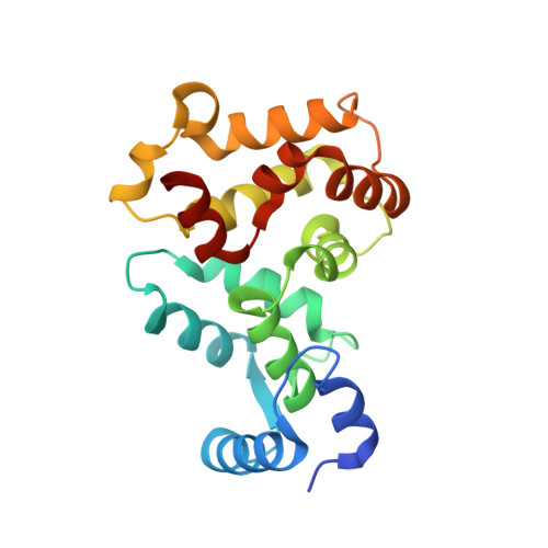
Crystal Structure of Recoverin with Calcium Ions Bound to Both Functional EF Hands.

Kumar, R.P.Ranaghan, M.J.Ganjei, A.Y.Oprian, D.D.

(2015) Biochemistry 54: 7222-7228

  • DOI: https://doi.org/10.1021/acs.biochem.5b01160
  • Primary Citation of Related Structures:  
    4YI8, 4YI9

  • PubMed Abstract: 

    Recoverin (Rv), a small Ca(2+)-binding protein that inhibits rhodopsin kinase (RK), has four EF hands, two of which are functional (EF2 and EF3). Activation requires Ca(2+) in both EF hands, but crystal structures have never been observed with Ca(2+) ions in both sites; all previous structures have Ca(2+) bound to only EF3. We suspected that this was due to an intermolecular crystal contact between T80 and a surface glutamate (E153) that precluded coordination of a Ca(2+) ion in EF2. We constructed the E153A mutant, determined its X-ray crystal structure to 1.2 Å resolution, and showed that two Ca(2+) ions are bound, one in EF3 and one in EF2. Additionally, several other residues are shown to adopt conformations in the 2Ca(2+) structure not seen previously and not seen in a second structure of the E153A mutant containing Na(+) instead of Ca(2+) in the EF2 site. The side-chain rearrangements in these residues form a 28 Å allosteric cascade along the surface of the protein connecting the Ca(2+)-binding site of EF2 with the active-site pocket responsible for binding RK.


  • Organizational Affiliation

    Department of Biochemistry, Brandeis University , Waltham, Massachusetts 02454, United States.


Macromolecules
Find similar proteins by:  (by identity cutoff)  |  3D Structure
Entity ID: 1
MoleculeChains Sequence LengthOrganismDetailsImage
Recoverin201Bos taurusMutation(s): 1 
Gene Names: RCVRNRCV1
UniProt
Find proteins for P21457 (Bos taurus)
Explore P21457 
Go to UniProtKB:  P21457
Entity Groups  
Sequence Clusters30% Identity50% Identity70% Identity90% Identity95% Identity100% Identity
UniProt GroupP21457
Sequence Annotations
Expand
  • Reference Sequence
Experimental Data & Validation

Experimental Data

  • Method: X-RAY DIFFRACTION
  • Resolution: 1.35 Å
  • R-Value Free: 0.174 
  • R-Value Work: 0.152 
  • R-Value Observed: 0.153 
  • Space Group: I 4
Unit Cell:
Length ( Å )Angle ( ˚ )
a = 85.34α = 90
b = 85.34β = 90
c = 58.75γ = 90
Software Package:
Software NamePurpose
MOSFLMdata reduction
SCALAdata scaling
PHASERphasing
PHENIXrefinement
PDB_EXTRACTdata extraction

Structure Validation

View Full Validation Report


Entry History & Funding Information

Deposition Data


Funding OrganizationLocationGrant Number
National Institutes of Health/National Institute of General Medical Sciences (NIH/NIGMS)United States--

Revision History  (Full details and data files)

  • Version 1.0: 2015-12-02
    Type: Initial release
  • Version 1.1: 2015-12-30
    Changes: Database references
  • Version 1.2: 2017-09-20
    Changes: Author supporting evidence, Database references, Derived calculations
  • Version 1.3: 2019-12-25
    Changes: Author supporting evidence
  • Version 1.4: 2023-09-27
    Changes: Data collection, Database references, Refinement description