4YAR

2-Hydroxyethylphosphonate dioxygenase (HEPD) E176H


Experimental Data Snapshot

  • Method: X-RAY DIFFRACTION
  • Resolution: 1.75 Å
  • R-Value Free: 0.238 
  • R-Value Work: 0.196 
  • R-Value Observed: 0.198 

wwPDB Validation   3D Report Full Report


This is version 1.4 of the entry. See complete history


Literature

A Common Late-Stage Intermediate in Catalysis by 2-Hydroxyethyl-phosphonate Dioxygenase and Methylphosphonate Synthase.

Peck, S.C.Chekan, J.R.Ulrich, E.C.Nair, S.K.van der Donk, W.A.

(2015) J Am Chem Soc 137: 3217-3220

  • DOI: https://doi.org/10.1021/jacs.5b00282
  • Primary Citation of Related Structures:  
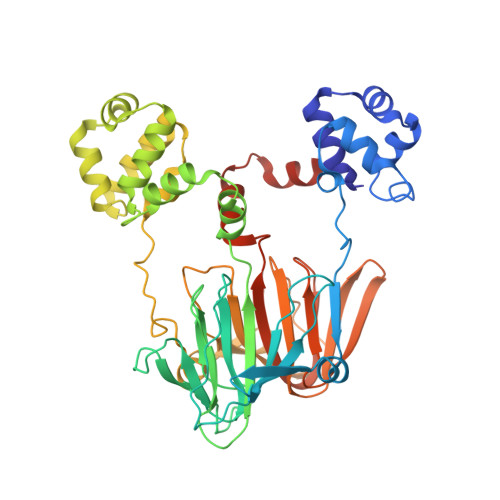
    4YAR

  • PubMed Abstract: 

    2-Hydroxyethylphosphonate dioxygenase (HEPD) and methylphosphonate synthase (MPnS) are nonheme iron oxygenases that both catalyze the carbon-carbon bond cleavage of 2-hydroxyethylphosphonate but generate different products. Substrate labeling experiments led to a mechanistic hypothesis in which the fate of a common intermediate determined product identity. We report here the generation of a bifunctional mutant of HEPD (E176H) that exhibits the activity of both HEPD and MPnS. The product distribution of the mutant is sensitive to a substrate isotope effect, consistent with an isotope-sensitive branching mechanism involving a common intermediate. The X-ray structure of the mutant was determined and suggested that the introduced histidine does not coordinate the active site metal, unlike the iron-binding glutamate it replaced.


  • Organizational Affiliation

    Howard Hughes Medical Institute and Department of Chemistry, University of Illinois at Urbana-Champaign , 600 South Mathews Avenue, Urbana, Illinois 61801, United States.


Macromolecules
Find similar proteins by:  (by identity cutoff)  |  3D Structure
Entity ID: 1
MoleculeChains Sequence LengthOrganismDetailsImage
2-hydroxyethylphosphonate dioxygenase443Streptomyces viridochromogenesMutation(s): 1 
Gene Names: hepDphpDSSQG_01041
EC: 1.13.11.72
UniProt
Find proteins for Q5IW40 (Streptomyces viridochromogenes (strain DSM 40736 / JCM 4977 / BCRC 1201 / Tue 494))
Explore Q5IW40 
Go to UniProtKB:  Q5IW40
Entity Groups  
Sequence Clusters30% Identity50% Identity70% Identity90% Identity95% Identity100% Identity
UniProt GroupQ5IW40
Sequence Annotations
Expand
  • Reference Sequence
Small Molecules
Ligands 3 Unique
IDChains Name / Formula / InChI Key2D Diagram3D Interactions
CD
Query on CD

Download Ideal Coordinates CCD File 
B [auth A]
C [auth A]
D [auth A]
E [auth A]
F [auth A]
B [auth A],
C [auth A],
D [auth A],
E [auth A],
F [auth A],
G [auth A],
H [auth A],
I [auth A],
J [auth A],
K [auth A]
CADMIUM ION
Cd
WLZRMCYVCSSEQC-UHFFFAOYSA-N
ACT
Query on ACT

Download Ideal Coordinates CCD File 
M [auth A],
N [auth A]
ACETATE ION
C2 H3 O2
QTBSBXVTEAMEQO-UHFFFAOYSA-M
NA
Query on NA

Download Ideal Coordinates CCD File 
L [auth A]SODIUM ION
Na
FKNQFGJONOIPTF-UHFFFAOYSA-N
Experimental Data & Validation

Experimental Data

  • Method: X-RAY DIFFRACTION
  • Resolution: 1.75 Å
  • R-Value Free: 0.238 
  • R-Value Work: 0.196 
  • R-Value Observed: 0.198 
  • Space Group: C 2 2 21
Unit Cell:
Length ( Å )Angle ( ˚ )
a = 85.286α = 90
b = 95.953β = 90
c = 101.142γ = 90
Software Package:
Software NamePurpose
REFMACrefinement
Aimlessdata scaling
PHASERphasing

Structure Validation

View Full Validation Report


Entry History 

Deposition Data

Revision History  (Full details and data files)

  • Version 1.0: 2015-03-04
    Type: Initial release
  • Version 1.1: 2015-03-25
    Changes: Database references
  • Version 1.2: 2015-10-14
    Changes: Data collection
  • Version 1.3: 2017-11-22
    Changes: Derived calculations, Refinement description
  • Version 1.4: 2023-09-27
    Changes: Data collection, Database references, Derived calculations, Refinement description