4Y7F

Crystal structure of glucosyl-3-phosphoglycerate synthase from Mycobacterium tuberculosis in complex with Mn2+, uridine-diphosphate-glucose (UDP-Glc) and 3-(phosphonooxy)propanoic acid (PPA) - GpgS Mn2+ UDP-Glc PPA


Experimental Data Snapshot

  • Method: X-RAY DIFFRACTION
  • Resolution: 3.23 Å
  • R-Value Free: 0.208 
  • R-Value Work: 0.170 
  • R-Value Observed: 0.172 

wwPDB Validation   3D Report Full Report


Ligand Structure Quality Assessment 


This is version 1.3 of the entry. See complete history


Literature

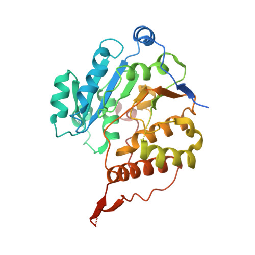
A Native Ternary Complex Trapped in a Crystal Reveals the Catalytic Mechanism of a Retaining Glycosyltransferase.

Albesa-Jove, D.Mendoza, F.Rodrigo-Unzueta, A.Gomollon-Bel, F.Cifuente, J.O.Urresti, S.Comino, N.Gomez, H.Romero-Garcia, J.Lluch, J.M.Sancho-Vaello, E.Biarnes, X.Planas, A.Merino, P.Masgrau, L.Guerin, M.E.

(2015) Angew Chem Int Ed Engl 54: 9898-9902

  • DOI: https://doi.org/10.1002/anie.201504617
  • Primary Citation of Related Structures:  
    4Y6N, 4Y6U, 4Y7F, 4Y7G, 4Y9X

  • PubMed Abstract: 

    Glycosyltransferases (GTs) comprise a prominent family of enzymes that play critical roles in a variety of cellular processes, including cell signaling, cell development, and host-pathogen interactions. Glycosyl transfer can proceed with either inversion or retention of the anomeric configuration with respect to the reaction substrates and products. The elucidation of the catalytic mechanism of retaining GTs remains a major challenge. A native ternary complex of a GT in a productive mode for catalysis is reported, that of the retaining glucosyl-3-phosphoglycerate synthase GpgS from M. tuberculosis in the presence of the sugar donor UDP-Glc, the acceptor substrate phosphoglycerate, and the divalent cation cofactor. Through a combination of structural, chemical, enzymatic, molecular dynamics, and quantum-mechanics/molecular-mechanics (QM/MM) calculations, the catalytic mechanism was unraveled, thereby providing a strong experimental support for a front-side substrate-assisted SN i-type reaction.


  • Organizational Affiliation

    Unidad de Biofísica, Consejo Superior de Investigaciones Científicas - Universidad del País Vasco/Euskal Herriko Unibertsitatea (CSIC-UPV/EHU) and Departamento de Bioquímica, Universidad del País Vasco, 48940 Leioa, Bizkaia (Spain).


Macromolecules
Find similar proteins by:  (by identity cutoff)  |  3D Structure
Entity ID: 1
MoleculeChains Sequence LengthOrganismDetailsImage
Glucosyl-3-phosphoglycerate synthase328Mycobacterium tuberculosis H37RvMutation(s): 0 
Gene Names: gpgSRv1208
EC: 2.4.1.266
UniProt
Find proteins for P9WMW9 (Mycobacterium tuberculosis (strain ATCC 25618 / H37Rv))
Explore P9WMW9 
Go to UniProtKB:  P9WMW9
Entity Groups  
Sequence Clusters30% Identity50% Identity70% Identity90% Identity95% Identity100% Identity
UniProt GroupP9WMW9
Sequence Annotations
Expand
  • Reference Sequence
Small Molecules
Ligands 5 Unique
IDChains Name / Formula / InChI Key2D Diagram3D Interactions
UPG
Query on UPG

Download Ideal Coordinates CCD File 
D [auth A]URIDINE-5'-DIPHOSPHATE-GLUCOSE
C15 H24 N2 O17 P2
HSCJRCZFDFQWRP-JZMIEXBBSA-N
48X
Query on 48X

Download Ideal Coordinates CCD File 
C [auth A]3-(phosphonooxy)propanoic acid
C3 H7 O6 P
QMMAAPSPIPKBBV-UHFFFAOYSA-N
SO4
Query on SO4

Download Ideal Coordinates CCD File 
E [auth A]SULFATE ION
O4 S
QAOWNCQODCNURD-UHFFFAOYSA-L
EDO
Query on EDO

Download Ideal Coordinates CCD File 
F [auth A]1,2-ETHANEDIOL
C2 H6 O2
LYCAIKOWRPUZTN-UHFFFAOYSA-N
MN
Query on MN

Download Ideal Coordinates CCD File 
B [auth A]MANGANESE (II) ION
Mn
WAEMQWOKJMHJLA-UHFFFAOYSA-N
Experimental Data & Validation

Experimental Data

  • Method: X-RAY DIFFRACTION
  • Resolution: 3.23 Å
  • R-Value Free: 0.208 
  • R-Value Work: 0.170 
  • R-Value Observed: 0.172 
  • Space Group: I 41
Unit Cell:
Length ( Å )Angle ( ˚ )
a = 97.94α = 90
b = 97.94β = 90
c = 126.78γ = 90
Software Package:
Software NamePurpose
PHENIXrefinement
XDSdata reduction
XSCALEdata scaling
Cootmodel building
PHASERphasing

Structure Validation

View Full Validation Report


Ligand Structure Quality Assessment 


Entry History 

Deposition Data

Revision History  (Full details and data files)

  • Version 1.0: 2015-07-15
    Type: Initial release
  • Version 1.1: 2015-08-19
    Changes: Database references
  • Version 1.2: 2016-01-27
    Changes: Structure summary
  • Version 1.3: 2024-01-10
    Changes: Data collection, Database references, Derived calculations, Refinement description