4XYJ

Crystal structure of human phosphofructokinase-1 in complex with ATP and Mg, Northeast Structural Genomics Consortium Target HR9275


Experimental Data Snapshot

  • Method: X-RAY DIFFRACTION
  • Resolution: 3.10 Å
  • R-Value Free: 0.246 
  • R-Value Work: 0.221 
  • R-Value Observed: 0.221 

wwPDB Validation   3D Report Full Report


Ligand Structure Quality Assessment 


This is version 1.5 of the entry. See complete history


Literature

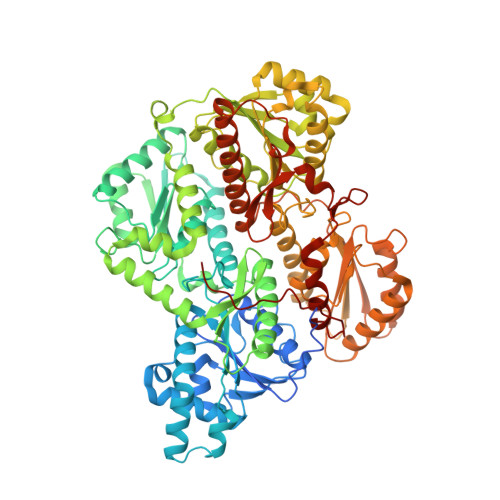
Structures of human phosphofructokinase-1 and atomic basis of cancer-associated mutations.

Webb, B.A.Forouhar, F.Szu, F.E.Seetharaman, J.Tong, L.Barber, D.L.

(2015) Nature 523: 111-114

  • DOI: https://doi.org/10.1038/nature14405
  • Primary Citation of Related Structures:  
    4XYJ, 4XYK

  • PubMed Abstract: 

    Phosphofructokinase-1 (PFK1), the 'gatekeeper' of glycolysis, catalyses the committed step of the glycolytic pathway by converting fructose-6-phosphate to fructose-1,6-bisphosphate. Allosteric activation and inhibition of PFK1 by over ten metabolites and in response to hormonal signalling fine-tune glycolytic flux to meet energy requirements. Mutations inhibiting PFK1 activity cause glycogen storage disease type VII, also known as Tarui disease, and mice deficient in muscle PFK1 have decreased fat stores. Additionally, PFK1 is proposed to have important roles in metabolic reprogramming in cancer. Despite its critical role in glucose flux, the biologically relevant crystal structure of the mammalian PFK1 tetramer has not been determined. Here we report the first structures of the mammalian PFK1 tetramer, for the human platelet isoform (PFKP), in complex with ATP-Mg(2+) and ADP at 3.1 and 3.4 Å, respectively. The structures reveal substantial conformational changes in the enzyme upon nucleotide hydrolysis as well as a unique tetramer interface. Mutations of residues in this interface can affect tetramer formation, enzyme catalysis and regulation, indicating the functional importance of the tetramer. With altered glycolytic flux being a hallmark of cancers, these new structures allow a molecular understanding of the functional consequences of somatic PFK1 mutations identified in human cancers. We characterize three of these mutations and show they have distinct effects on allosteric regulation of PFKP activity and lactate production. The PFKP structural blueprint for somatic mutations as well as the catalytic site can guide therapeutic targeting of PFK1 activity to control dysregulated glycolysis in disease.


  • Organizational Affiliation

    Department of Cell and Tissue Biology, University of California, San Francisco, California 94143, USA.


Macromolecules
Find similar proteins by:  (by identity cutoff)  |  3D Structure
Entity ID: 1
MoleculeChains Sequence LengthOrganismDetailsImage
ATP-dependent 6-phosphofructokinase, platelet type
A, B, C, D, E
A, B, C, D, E, F, G, H
812Homo sapiensMutation(s): 0 
Gene Names: PFKPPFKF
EC: 2.7.1.11
UniProt & NIH Common Fund Data Resources
Find proteins for Q01813 (Homo sapiens)
Explore Q01813 
Go to UniProtKB:  Q01813
PHAROS:  Q01813
GTEx:  ENSG00000067057 
Entity Groups  
Sequence Clusters30% Identity50% Identity70% Identity90% Identity95% Identity100% Identity
UniProt GroupQ01813
Sequence Annotations
Expand
  • Reference Sequence
Small Molecules
Ligands 4 Unique
IDChains Name / Formula / InChI Key2D Diagram3D Interactions
ATP
Query on ATP

Download Ideal Coordinates CCD File 
AA [auth E]
EA [auth F]
I [auth A]
KA [auth G]
N [auth B]
AA [auth E],
EA [auth F],
I [auth A],
KA [auth G],
N [auth B],
PA [auth H],
T [auth C],
X [auth D]
ADENOSINE-5'-TRIPHOSPHATE
C10 H16 N5 O13 P3
ZKHQWZAMYRWXGA-KQYNXXCUSA-N
PO4
Query on PO4

Download Ideal Coordinates CCD File 
DA [auth E]
HA [auth F]
IA [auth F]
L [auth A]
M [auth A]
DA [auth E],
HA [auth F],
IA [auth F],
L [auth A],
M [auth A],
NA [auth G],
OA [auth G],
Q [auth B],
R [auth B],
SA [auth H],
W [auth C],
Z [auth D]
PHOSPHATE ION
O4 P
NBIIXXVUZAFLBC-UHFFFAOYSA-K
CO
Query on CO

Download Ideal Coordinates CCD File 
JA [auth F],
S [auth B]
COBALT (II) ION
Co
XLJKHNWPARRRJB-UHFFFAOYSA-N
MG
Query on MG

Download Ideal Coordinates CCD File 
BA [auth E]
CA [auth E]
FA [auth F]
GA [auth F]
J [auth A]
BA [auth E],
CA [auth E],
FA [auth F],
GA [auth F],
J [auth A],
K [auth A],
LA [auth G],
MA [auth G],
O [auth B],
P [auth B],
QA [auth H],
RA [auth H],
U [auth C],
V [auth C],
Y [auth D]
MAGNESIUM ION
Mg
JLVVSXFLKOJNIY-UHFFFAOYSA-N
Experimental Data & Validation

Experimental Data

  • Method: X-RAY DIFFRACTION
  • Resolution: 3.10 Å
  • R-Value Free: 0.246 
  • R-Value Work: 0.221 
  • R-Value Observed: 0.221 
  • Space Group: P 1 21 1
Unit Cell:
Length ( Å )Angle ( ˚ )
a = 137.199α = 90
b = 159.329β = 104.2
c = 170.533γ = 90
Software Package:
Software NamePurpose
CNSrefinement
HKL-2000data reduction
SCALEPACKdata scaling
MOLREPphasing

Structure Validation

View Full Validation Report


Ligand Structure Quality Assessment 


Entry History 

Deposition Data

Revision History  (Full details and data files)

  • Version 1.0: 2015-05-20
    Type: Initial release
  • Version 1.1: 2015-06-03
    Changes: Database references
  • Version 1.2: 2015-07-15
    Changes: Database references
  • Version 1.3: 2017-11-01
    Changes: Author supporting evidence, Database references, Derived calculations, Source and taxonomy
  • Version 1.4: 2019-11-20
    Changes: Derived calculations
  • Version 1.5: 2023-09-27
    Changes: Data collection, Database references, Derived calculations, Refinement description